| Journal of Clinical Medicine Research, ISSN 1918-3003 print, 1918-3011 online, Open Access |
| Article copyright, the authors; Journal compilation copyright, J Clin Med Res and Elmer Press Inc |
| Journal website https://jocmr.elmerjournals.com |
Original Article
Volume 18, Number 9, September 2026, pages 652-660

Common Carotid Artery Extra-Media Thickness as a Marker of Perivascular Obesity and Cardiometabolic Risk in Individuals Without Cardiovascular Disease
Anna Braginaa  , Natalia Druzhininaa, d , Yulia Rodionovaa , Daria Akvitskayaa , Kirill Novikova , Galina Nikiforovab , Oksana Avdeenkoc , Natalia Vlasovac , Alina Arzukanyanc , Alina Kudravcevaa , Valery Podzolkova
aDepartment of Internal Medicine No. 2, Institute of Clinical Medicine, Sechenov First Moscow State Medical University, 19991 Moscow, Russia
bDepartment for Ear, Nose and Throat Diseases, Institute of Clinical Medicine, Sechenov First Moscow State Medical University, 19991 Moscow, Russia
cDepartment of Therapeutic Dentistry, Institute of Dentistry Named After E.V. Borovsky, Sechenov First Moscow State Medical University, 19991 Moscow, Russia
dCorresponding Author: Natalia Druzhinina, Department of Internal Medicine No. 2, Institute of Clinical Medicine, Sechenov First Moscow State Medical University, 19991Moscow, Russia
Manuscript submitted July 13, 2026, accepted September 3, 2026, published online September 26, 2026
Short title: Carotid Artery Thickness and Risk Factors
doi: https://doi.org/10.14740/jocmr6657
| Abstract | ▴Top |
Background: Perivascular adipose tissue (PVAT) is increasingly recognized as a pathogenetic contributor to vascular dysfunction. Carotid extra-media thickness (EMT) measured by ultrasound is the simplest, noninvasive and clinically accessible marker of PVAT deposition. However, reference values in young individuals without cardiovascular disease (CVD) are lacking. The aim of this study was to analyze the relationship between carotid EMT and cardiovascular risk factors and to determine a tentative reference value of carotid EMT in individuals without CVD.
Methods: This cross-sectional study included 373 patients with a median age of 23 (21; 32) years and without CVD. All patients underwent anthropometric examination, lipid-profile assessment using the CardioChek PA (USA, 2020), and duplex ultrasound of the brachiocephalic arteries with measurement of common carotid artery EMT and intima-media thickness (IMT). A subgroup of apparently healthy individuals without cardiovascular risk factors was selected to define a threshold. EMT above the 90th percentile of this subgroup was regarded as perivascular obesity (PVO).
Results: Carotid EMT was significantly associated with age, blood pressure levels, anthropometric and laboratory metabolic markers, glycemia and smoking. On multivariable linear regression, age, obesity and smoking were independently associated with EMT. The 90th percentile threshold in the apparently healthy subgroup (n = 185) was 0.54 mm; EMT ≥ 0.54 mm (PVO) was identified in 24.2% of the sample and was most frequent in individuals aged 41–55 years. On multivariable logistic regression, age and waist circumference were independently associated with the presence of PVO.
Conclusions: Carotid EMT, reflecting the severity of PVAT deposition, may be associated with a wide range of cardiometabolic risk factors in individuals without CVD, with a tentative threshold value of 0.54 mm.
Keywords: Adipose tissue; Extra-media thickness; Perivascular obesity; Carotid arteries; Obesity; Risk factors
| Introduction | ▴Top |
Obesity represents a significant medical and social problem worldwide [1], with a high prevalence not only in older adults [2] but also in younger age groups [3, 4]. Although the rate of increase in obesity prevalence has slowed in several high-income countries [5], the global burden of obesity and associated cardiovascular complications continues to rise [2].
Obesity is characterized by a variety of phenotypes [6]. Some of these phenotypes may evolve over time into a metabolically unhealthy phenotype with increased cardiovascular risk [7]. The variability of adipose tissue distribution is well recognized and is partly related to the formation of ectopic fat depots [8–10]. Various visceral fat depots, including pararenal adipose tissue (PRAT) [11], pericardial adipose tissue (PAT) [12], and perivascular adipose tissue (PVAT) [13], are considered potential contributors to dysfunction of the organs they surround [14]. With excessive accumulation, PVAT may develop a dysfunctional phenotype and synthesize a broad spectrum of adipokines that contribute to meta-inflammation, vascular remodeling, increased vascular stiffness, atherosclerosis, and thrombosis [13–15].
For PVAT imaging, several consensus documents recommend ultrasound, magnetic resonance imaging, multislice computed tomography (MSCT), and positron emission tomography-computed tomography [16]. We have previously proposed tentative reference values for periaortic adipose tissue based on chest MSCT data [17]. However, the use of radiological methods may be limited by radiation exposure, cost, and accessibility. In this regard, ultrasound of the brachiocephalic arteries may be considered a clinically accessible approach for evaluating PVAT. Ultrasound measurement of common carotid artery extra-media thickness (EMT) has been proposed as a simple, noninvasive, and clinically available marker of PVAT [17]. The extra-media complex comprises the arterial adventitia and the PVAT surrounding the vessel [18].
An association of EMT with obesity and metabolic syndrome (MS) has been demonstrated in elderly patients [19]. It has been suggested that EMT, unlike intima-media thickness (IMT), may reflect not only vascular wall stiffness but also PVAT accumulation as a potential pathogenetic contributor to vascular dysfunction [19]. A meta-analysis demonstrated a significant relationship between PVAT, assessed by ultrasound and radiological methods, and hypertension, dyslipidemia, and obesity markers, including body mass index (BMI), waist circumference (WC) [20, 21]. Nevertheless, data on reference values of carotid EMT as an accessible marker of PVAT in young individuals without cardiovascular disease (CVD) remain limited.
Accordingly, the aim of our study was to analyze the relationship between carotid EMT and cardiovascular risk factors and to determine a tentative reference value of carotid EMT in individuals without CVD.
| Materials and Methods | ▴Top |
This cross-sectional study was conducted at University Clinical Hospital No. 4 of Sechenov University in accordance with the ethical principles of the Declaration of Helsinki. The study was approved by the local ethics committee on December 8, 2022 (protocol No. 25–22). All participants provided written informed consent. The inclusion criteria were an age between 18 and 55 years and consent to participate in the study.
The exclusion criteria were as follows: presence of symptomatic hypertension; clinical manifestations of CVD, including coronary heart disease and cerebrovascular disease; presence of atherosclerotic plaques; clinical and laboratory manifestations of chronic liver disease; decreased glomerular filtration rate < 60 mL/min/1.73 m2; proteinuria ≥ 300 mg/day; type 1 or type 2 diabetes mellitus; inflammatory or oncological diseases of any localization; pregnancy at the time of inclusion; and use of medications for the treatment of obesity or dyslipidemia.
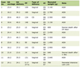
The study included 373 patients with a median age of 23 (21; 32) years (Table 1). History data on the presence and duration of hypertension and smoking status were collected. All patients underwent anthropometric examination with measurement of WC, hip circumference (HC), and neck circumference. BMI was calculated as weight divided by height squared (kg/m2). Biochemical lipid-profile parameters, including total cholesterol (TC), low-density lipoprotein cholesterol (LDL-C), high-density lipoprotein cholesterol (HDL-C), and triglycerides (TG), were assessed using a CardioChek PA express analyzer (USA, 2020). Overweight and obesity grade were assessed using BMI in accordance with clinical guidelines [22] and by WC [23]. Abdominal obesity (AO) was diagnosed when WC was > 102 cm in men and > 88 cm in women [23]. The hypertension grade was defined in accordance with the 2023 guidelines of the European Society of Cardiology (ESC)/European Society of Hypertension [24]. Dyslipidemia was defined according to the ESC/European Atherosclerosis Society guidelines for the management of dyslipidemias: lipid modification to reduce cardiovascular risk, 2024 [25]. Diabetes mellitus and impaired fasting glycemia were defined in accordance with the 2022 guidelines of the Russian Association of Endocrinologists for type 2 diabetes mellitus in adults. We assessed impaired fasting glycemia based on questionnaire data and the interpretation of laboratory test results according to the following threshold values: fasting venous blood glucose < 7.0 mmol/L and (if performed) post-oral glucose tolerance test < 7.8 mmol/L [26].
 Click to view | Table 1. Clinical Characteristics of the Patients Included in the Study |
Assessment of the brachiocephalic arteries and measurement of common carotid artery EMT were performed by duplex ultrasound using a Philips Epiq 7DS system (USA, 2018) according to the method described by Skilton et al [27]. Carotid EMT was assessed with a linear transducer at a point 1–1.5 cm proximal to the common carotid artery bifurcation. Carotid EMT was defined as the distance between the inner surface of the posterior wall of the internal jugular vein (relative to the transducer) and the outer surface of the anterior wall of the common carotid artery [27].
IMT was assessed by duplex ultrasound of the brachiocephalic arteries. The results were interpreted as follows: IMT < 0.9 mm was considered normal; 0.9 ≤ IMT < 1.5 mm was considered subclinical atherosclerosis; and IMT ≥ 1.5 mm or focal thickening of 0.5 mm compared with IMT values in adjacent carotid artery segments was considered an atherosclerotic plaque. Patients with atherosclerotic plaques were not included in the study. Measurement of carotid IMT, as well as EMT, was performed bilaterally. Thickness measurements on each side were taken three times, and the mean value was recorded in the study protocol and subsequently used for statistical analysis.
Statistical analysis
Statistical analysis of the results was performed using Jamovi (version 2.3) software. Variables with a non-normal distribution were presented as the median and the 25th and 75th percentiles. Variables with a normal distribution were presented as the mean with standard deviation. The significance of differences between continuous values was assessed using the Mann–Whitney U test (P (U)). Categorical variables were compared using Pearson’s chi-square test (P (χ2)). Spearman’s rank correlation coefficient (ρ) was used to evaluate associations between the studied parameters. Multivariable linear regression analysis was used to assess independent associations between selected predictors and continuous dependent variables, whereas logistic regression analysis was used for binary dependent variables.
For multivariate logistic regression analysis, all continuous independent variables were standardized using the z-transform to obtain an average value of 0 and a standard deviation of 1. Thus, the odds ratios (ORs) represent the change in the probability of developing perivascular obesity (PVO) by the amount of an increase in the corresponding variable by one standard deviation.
To determine the threshold value of EMT, a subgroup of apparently healthy individuals without cardiovascular risk factors, including obesity, hypertension, dyslipidemia, and impaired glucose tolerance (IGT), and impaired fasting glycemia, was selected from the total sample. EMT values exceeding the 90th percentile of the distribution in this subgroup were considered indicative of excessive PVAT deposition, hereafter termed PVO, whereas values below this threshold were regarded as normal EMT.
| Results | ▴Top |
The clinical characteristics of the examined sample, including the prevalence of cardiovascular risk factors such as obesity according to BMI, AO, hypertension, smoking, dyslipidemia, and impaired fasting glycemia, as well as the median IMT and EMT of the common carotid arteries, are presented in Table 1.
In the total study sample, the median carotid EMT was 0.43 (0.336; 0.535) mm, whereas the median carotid IMT was 0.70 (0.63; 0.85) mm (Table 1). Both EMT and IMT of the carotid arteries showed an abnormal distribution (Shapiro–Wilk test: P = 0.004 and P < 0.001, respectively) (Fig. 1).
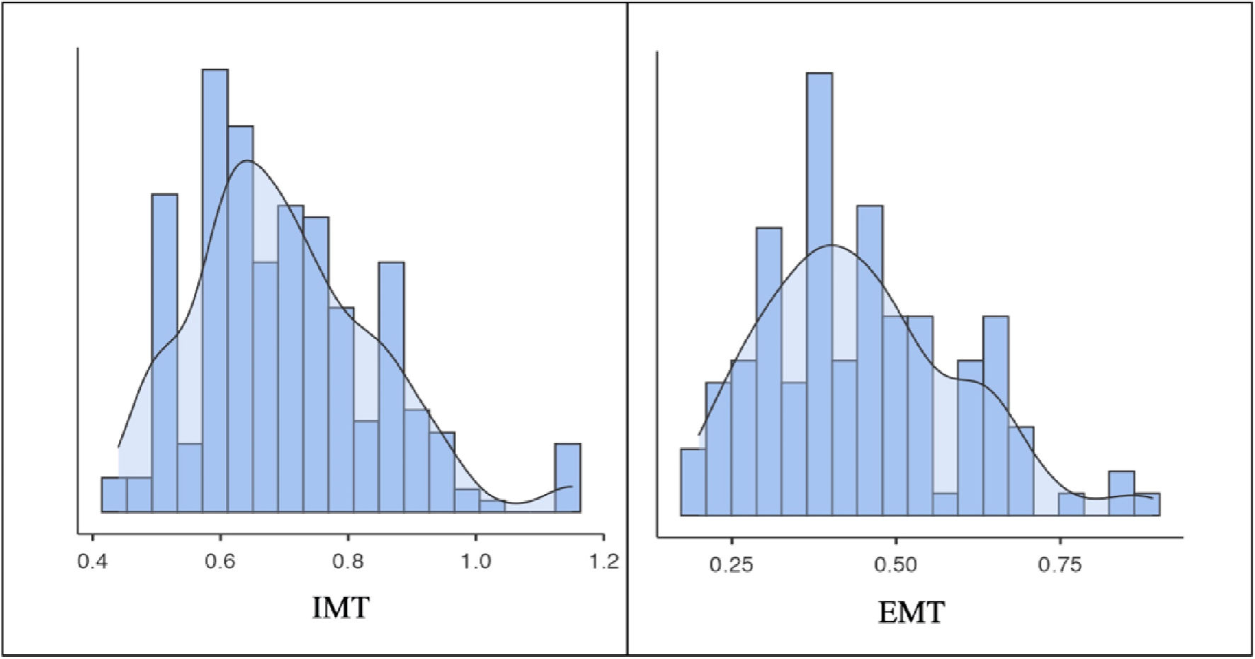 Click for large image | Figure 1. Distribution of the IMT and the EMT of the carotid arteries. EMT: common carotid artery extra-media thickness; IMT: intima-media thickness. |
To assess the relationship between carotid EMT and cardiovascular risk factors, univariate correlation analysis was performed. The results are presented in Figure 2.
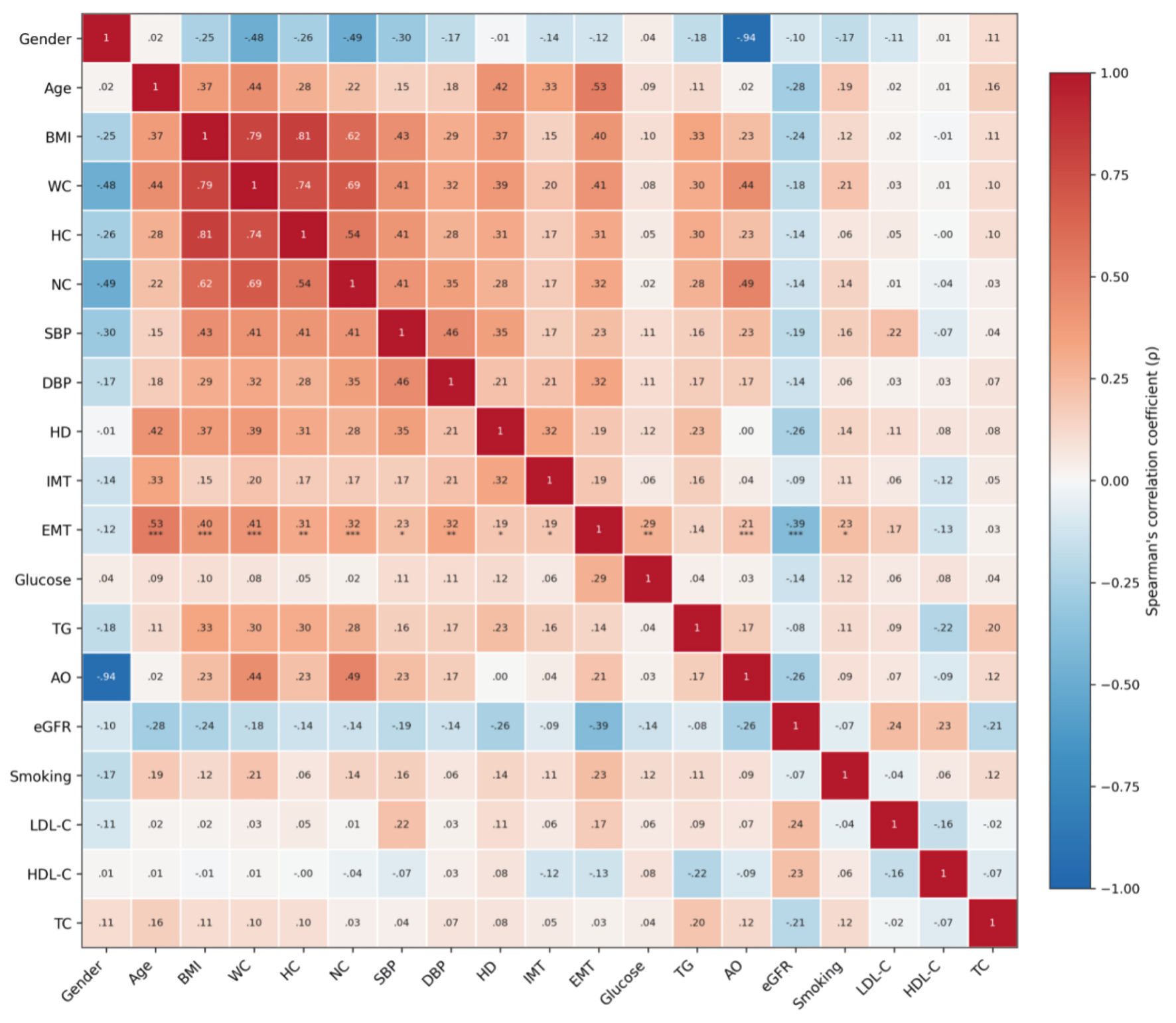 Click for large image | Figure 2. Correlation matrix (heat map) in the examined sample. *P < 0.05; **P < 0.01; ***P < 0.001. AO: abdominal obesity; BMI: body mass index; DBP: diastolic blood pressure; eGFR: estimated glomerular filtration rate; EMT: common carotid artery extra-media thickness; HC: hip circumference; HD: hypertension duration; HDL-C: high-density lipoprotein cholesterol; IMT: intima-media thickness; LDL-C: low-density lipoprotein cholesterol; NC: neck circumference; SBP: systolic blood pressure; TC: total cholesterol; TG: triglycerides; WC: waist circumference. |
To assess the independent associations between various factors and EMT, a multivariable linear regression analysis was performed. Carotid EMT was specified as the continuous dependent variable. Models were constructed sequentially. Traditional risk factors (age, sex, obesity, hypertension, smoking, dyslipidemia, impaired fasting glycemia), cardiometabolic risk factors (hypertension duration, smoking duration, blood pressure, BMI, WC, and HC), and biochemical parameters (glycemia, TC, TG, LDL-C, and HDL-C levels) were considered as independent variables. The model with the highest statistical significance is presented in Table 2.
Click to view | Table 2. Multivariable Linear Regression Analysis With Carotid EMT as the Dependent Variable, Adjusted for Age, Obesity, Hypertension, and Smoking |
To determine a tentative EMT threshold, a subgroup of individuals without cardiovascular risk factors (obesity, hypertension, dyslipidemia, impaired fasting glycemia, and smoking) was selected from the total sample (n = 373). This apparently healthy subgroup comprised 185 individuals, accounting for 49.6% of the total sample. The threshold was defined as the 90th percentile of the EMT distribution in this subgroup. Accordingly, EMT < 0.54 mm was regarded as normal, whereas EMT ≥ 0.54 mm was used as an operational criterion for identifying PVO. Based on this criterion, PVO was identified in 24.2% of patients in the study sample.
PVO was most frequently observed in the group aged 41–55 years and significantly exceeded the prevalence of both BMI-defined obesity and AO (P < 0.001) (Fig. 3).
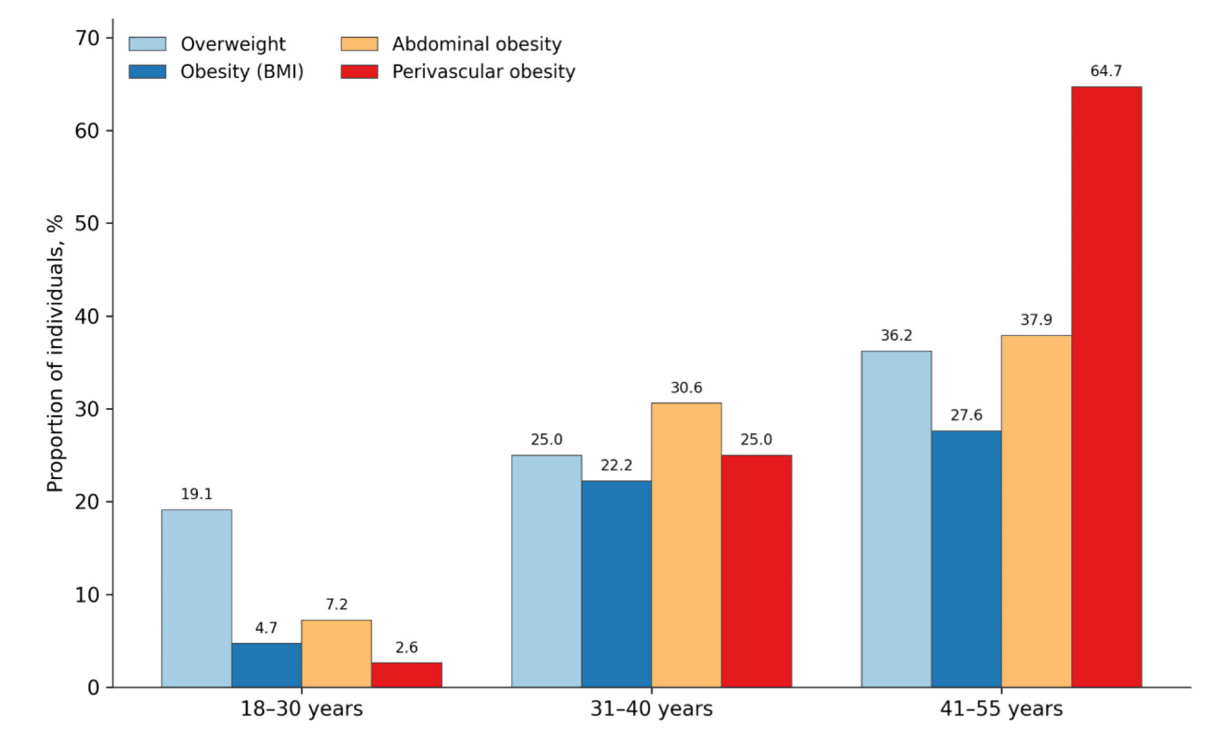 Click for large image | Figure 3. Proportion of individuals with perivascular obesity, overweight, obesity by BMI, and abdominal obesity, by age group. BMI: body mass index. |
PVO was detected significantly more often in individuals with obesity defined by BMI and in those with AO than in overweight individuals (P < 0.05) (Fig. 4). Notably, PVO was also identified in individuals with normal body weight (12.9%), suggesting that carotid EMT may identify PVAT accumulation not captured by conventional anthropometric criteria.
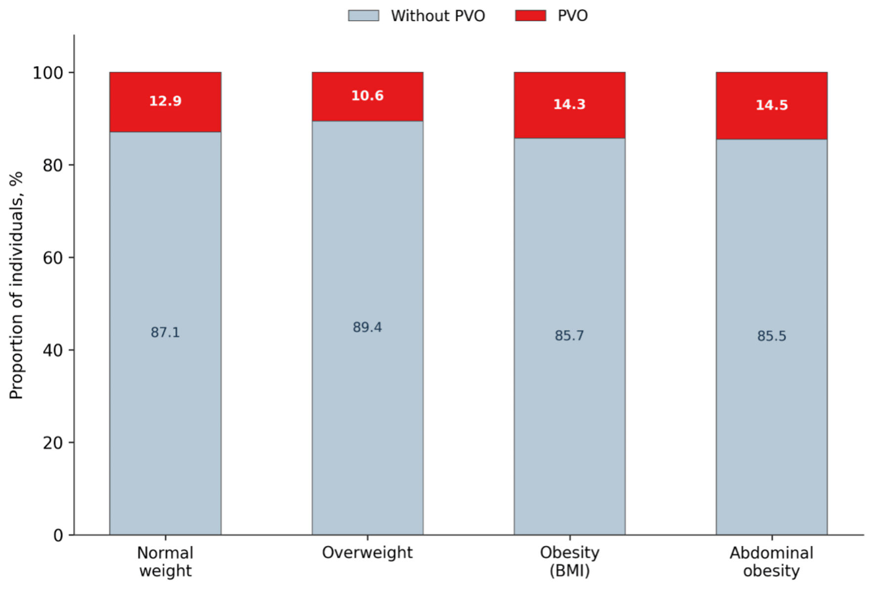 Click for large image | Figure 4. Frequency of individuals with perivascular obesity across groups with normal weight, overweight, obesity by BMI, and abdominal obesity in the examined sample. BMI: body mass index; PVO: perivascular obesity. |
To assess independent associations between various CVD risk factors and the presence of PVO, multivariable logistic regression analysis was performed. All continuous independent variables were standardized before multivariable logistic regression analysis. CVD risk factors and their anamnestic characteristics (duration), laboratory characteristics (dyslipidemia and glycemia markers), and instrumental characteristics (systolic blood pressure (SBP), diastolic blood pressure (DBP), and IMT) were sequentially included in the logistic regression models. The final model is presented in Table 3. Age and WC were independently associated with the presence of PVO. A 1-standard-deviation increase in age was associated with higher odds of PVO (OR = 5.22; 95% confidence interval (CI), 2.35–11.55; P < 0.001), as was a 1-standard-deviation increase in WC (OR = 2.82; 95% CI, 1.31–6.09; P = 0.008). Carotid IMT, hypertension duration, TC, and glucose were not independently associated with PVO (Table 3). According to this analysis, age and WC were independently associated with the presence of PVO among individuals without CVD.
Click to view | Table 3. Results of Multivariable Logistic Regression Analysis of the Association of Perivascular Obesity With Risk Factors Using Standardized Continuous Variables |
| Discussion | ▴Top |
Carotid EMT is a simple, noninvasive, and clinically accessible ultrasound-based approach for assessing PVAT [28, 29]. Standard examination protocols usually include blood-flow velocity parameters, vessel geometry, IMT, and, when present, the degree of stenosis [30]. In 2009, Skilton et al proposed a method for assessing carotid EMT, which has since been used as an ultrasound marker of PVAT [27].
According to several studies [28, 31, 32], carotid EMT is associated with age-related factors, including the degree of carotid atherosclerotic involvement [29], coronary heart disease [33], and cerebrovascular disease [34].
Our results demonstrated significant associations between carotid EMT and age, blood pressure, anthropometric parameters, laboratory metabolic markers, glycemia, and smoking (Fig. 1). In multivariable analysis, age, obesity, and smoking remained independently associated with EMT (Table 2). No independent association was found between hypertension and EMT, possibly because of the relatively small proportion of participants with hypertension (15%) and the short duration of hypertension in this subgroup.
Lefferts et al demonstrated that carotid EMT was associated not only with markers of visceral adiposity, including WC and sagittal abdominal diameter, but also with vascular stiffness [35]. Unlike their study, which included 135 healthy young men with a mean age of 20 ± 2 years, our study included both men and women across young and middle-aged groups. Similar findings were reported by Carlini et al, who demonstrated significant associations of EMT with age and SBP in a relatively small sample of participants (n = 50; mean age, 42.0 ± 19.0 years) [18].
The adventitial layer is considered a major contributor to increased EMT [36]. The arterial adventitia is a structurally and functionally complex component of the vessel wall that participates in the release and activation of signaling mediators involved in the regulation of vascular tone and homeostasis [37]. Age-related adventitial fibrosis and calcification have been suggested to contribute to the development of arterial stiffness [38]. In addition, dysfunctional PVAT may promote meta-inflammation, impair regional vascular tone and vasa vasorum perfusion, and contribute to increased vascular wall stiffness [37].
According to published data, carotid EMT measured using the method described by Skilton et al [27] was 0.63 mm in individuals with carotid atherosclerosis, 0.38 mm in middle-aged individuals with normal carotid anatomy [31], and 0.80 mm in patients with coronary heart disease [33]. In our study, a tentative EMT threshold of 0.54 mm was identified for increased PVAT in young and middle-aged individuals without CVD. The presence of PVO, defined as EMT ≥ 0.54 mm, was independently associated with age and WC.
Other studies have also demonstrated associations of EMT with AO and its anthropometric marker, WC, as well as with other ectopic fat depots [31]. Druzhilov et al found significant correlations of EMT with both AO and epicardial adipose tissue (r = 0.71, P < 0.001) [31]. In another study involving 100 patients, carotid EMT was associated with epicardial adipose tissue (r = 0.46, P < 0.001) and PAT (r = 0.30, P < 0.001); the mean carotid EMT in that study was 0.66 mm [19].
Our findings suggest that carotid EMT of 0.54 mm or greater may be considered a tentative criterion for identifying PVO. This threshold is within the range of mean EMT values previously reported across different study populations (0.3–0.7 mm) [19, 31, 33]. Further validation of the proposed threshold in various clinical subgroups (including males and females of different age groups, as well as individuals with anthropometric peculiarities, e.g., chest wall deformities [39]) may facilitate the development of novel diagnostic algorithms for stratifying patients according to their risk of CVD and related complications.
Among the limitations of this study, the predominance of young individuals should be noted: 272 participants (73% of the total sample) were younger than 30 years. Owing to the limited sample size, sex- and age-specific differences in tentative EMT reference values as markers of PVO were not assessed. Further studies with larger and more age-balanced samples are required to address these differences.
In conclusion, despite these limitations, carotid EMT was associated with a broad range of cardiometabolic risk factors in individuals without CVD and may reflect PVAT deposition. A tentative threshold of 0.54 mm was identified for detecting possible PVO.
Acknowledgments
None to declare.
Financial Disclosure
None to declare.
Conflict of Interest
The authors declare that they have no conflict of interest.
Informed Consent
All patients provided written voluntary informed consent prior to inclusion in the study.
Author Contributions
Conceptualization: AB, NV, YR, VP, GN; data curation: ND, YR, DA, KN, NV; formal analysis: ND, YR, DA, OA, AA; investigation: DA, OA, NV, AK; methodology: ND, YR, DA, AA; project administration: AB, NV, YR; resources: ND, OA, NV, AA, GN; software: ND, YR, DA, AK; supervision: AB, NV, YR; validation: ND, YR, DA, NV; visualization: NV, DA, AK; writing – original draft: NV, DA, KN, AK; writing – review and editing: AB, ND, YR, VP, GN. All authors approved the final version of the manuscript for submission.
Data Availability
The data that support the findings of this study are available from the corresponding author upon reasonable request.
AI Use Declaration
The authors confirm that generative artificial intelligence tools (ChatGPT, OpenAI) were used solely for language editing and to enhance the clarity and readability of the manuscript. These tools were not used for study design, data analysis, interpretation of results, or the development of scientific content, figures, or images. All scientific content, interpretations, and conclusions presented in the manuscript remain entirely the responsibility of the authors.
Abbreviations
AO: abdominal obesity; BMI: body mass index; CVD: cardiovascular disease; DBP: diastolic blood pressure; EMT: extra-media thickness; GFR: glomerular filtration rate; HC: hip circumference; HDL-C: high-density lipoprotein cholesterol; IMT: intima-media thickness; LDL-C: low-density lipoprotein cholesterol; MS: metabolic syndrome; MSCT: multislice computed tomography; NC: neck circumference; PAT: pericardial adipose tissue; PRAT: pararenal adipose tissue; PVAT: perivascular adipose tissue; PVO: perivascular obesity; SBP: systolic blood pressure; TC: total cholesterol; TG: triglycerides; WC: waist circumference
| References | ▴Top |
- World Health Organization. Obesity and overweight. https://www.who.int/news-room/fact-sheets/detail/obesity-and-overweight. Accessed January 18, 2025.
- NCD Risk Factor Collaboration. Worldwide trends in underweight and obesity from 1990 to 2022: a pooled analysis of 3663 population-representative studies with 222 million children, adolescents, and adults. Lancet. 2024;403(10431):1027-1050.
doi pubmed - Kansra AR, Lakkunarajah S, Jay MS. Childhood and adolescent obesity: a review. Front Pediatr. 2020;8:581461.
doi pubmed - Haseler E, Sinha MD. Tackling the epidemic of obesity and hypertension in children and young people. Curr Opin Nephrol Hypertens. 2026;35(2):156-163.
doi pubmed - Koliaki C, Dalamaga M, Liatis S. Update on the obesity epidemic: after the sudden rise, Is the upward trajectory beginning to flatten? Curr Obes Rep. 2023;12(4):514-527.
doi pubmed - Schulze MB. Metabolic health in normal-weight and obese individuals. Diabetologia. 2019;62(4):558-566.
doi pubmed - Kramer CK, Zinman B, Retnakaran R. Are metabolically healthy overweight and obesity benign conditions? A systematic review and meta-analysis. Ann Intern Med. 2013;159(11):758-769.
doi pubmed - Eckel N, Li Y, Kuxhaus O, Stefan N, Hu FB, Schulze MB. Transition from metabolic healthy to unhealthy phenotypes and association with cardiovascular disease risk across BMI categories in 90 257 women (the Nurses' Health Study): 30 year follow-up from a prospective cohort study. Lancet Diabetes Endocrinol. 2018;6(9):714-724.
doi pubmed - Podzolkov VI, Bragina AE, Vasilchenko MK, Osadchiy KK, Pilipenko MO, Antonenko VA, Mansimova LB, et al. Phenotypic clusters of obesity: gender, clinical, laboratory and genetic characteristics. Ter Arkh. 2025;97(9):773-780.
- Podzolkov VI, Bragina AE, Rodionova YN, Osadchiy KK, Vasilchenko MK, Muravlev AP, Ananeva VS, et al. Ectopic adipose tissue: frequency and clinical characteristics of obesity phenotypes in patients. Cardiovasc Ther Prev. 2024;23(6):50-56.
- Bragina capital A CIEC, Osadchiy KK, Rodionova JN, Bayutina Dcapital A C, Cherepanov capital A C, Podzolkov VI. Pararenal fat and renal dysfunction in patients without significant cardiovascular disease. Am J Nephrol. 2022;53(5):416-422.
doi pubmed - Gritsenko OV, Chumakova GA, Trubina EV. [The role of epicardial obesity in the development of left ventricular diastolic dysfunction]. Kardiologiia. 2023;63(7):32-38.
doi pubmed - Podzolkov VI, Bragina AE, Osadchiy KK, Rodionova YN, Jafarova ZB, Lobanova MV, Larionova YS. Relationship between the volume of perivascular adipose tissue and the vascular wall lesion. Cardiovasc Ther Prev. 2021;20(7):2993.
- Koskinas KC, Van Craenenbroeck EM, Antoniades C, Bluher M, Gorter TM, Hanssen H, Marx N, et al. Obesity and cardiovascular disease: an ESC clinical consensus statement. Eur Heart J. 2024;45(38):4063-4098.
doi pubmed - Bailey-Downs LC, Tucsek Z, Toth P, Sosnowska D, Gautam T, Sonntag WE, Csiszar A, et al. Aging exacerbates obesity-induced oxidative stress and inflammation in perivascular adipose tissue in mice: a paracrine mechanism contributing to vascular redox dysregulation and inflammation. J Gerontol A Biol Sci Med Sci. 2013;68(7):780-792.
doi pubmed - Foran D, Chan K, Antoniades C. Imaging and quantification of perivascular adipose tissue. Arterioscler Thromb Vasc Biol. 2026;46(3):e321704.
doi pubmed - Podzolkov VI, Bragina AE, Osadchiy KK, Rodionova YN, Djafarova ZB, Khalenyan MH, Dishkaya SO. [Ectopic obesity in patients without manifested cardiovascular disease: regulations, frequency and clinical characteristics]. Ter Arkh. 2022;94(9):1072-1077.
doi pubmed - Carlini NA, Harber MP, Fleenor BS. Age-related carotid extra-media thickening is associated with increased blood pressure and arterial stiffness. Clin Physiol Funct Imaging. 2021;41(5):461-466.
doi pubmed - Haberka M, Sosnowski M, Zuziak-Pruska J, Gasior Z. Extra-media thickness and epicardial fat: Comparison of a novel carotid artery ultrasound index and a well-established cardiac magnetic resonance fat quantification method. Nutr Metab Cardiovasc Dis. 2015;25(8):763-770.
doi pubmed - Bragina A, Rodionova Y, Druzhinina N, Suvorov A, Osadchiy K, Ishina T, Vasilchenko M, et al. Relationship between perivascular adipose tissue and cardiovascular risk factors: a systematic review and meta-analysis. Metab Syndr Relat Disord. 2024;22(1):1-14.
doi pubmed - Podzolkov VI, Safronova TA, Vasyuk YA, Kotovskaya YV, Kislyak OA, Starodubova AV, Kopylov FY, et al. Diagnosis of arterial stiffness using the cardio-ankle vascular index. Expert consensus. Cardiovasc Ther Prev. 2025;24(8):4481.
- Dedov II, Mokrysheva NG, Melnichenko GA, Mazurina NV, Andreeva EN, Bondarenko IZ, Gusova ZR, et al. Obesity. Clinical guidelines. Consilium Medicum. 2021;23(4):311-325.
- Nam GE, Park HS. Perspective on diagnostic criteria for obesity and abdominal obesity in Korean adults. J Obes Metab Syndr. 2018;27(3):134-142.
doi pubmed - Williams B, Mancia G, Spiering W, Agabiti Rosei E, Azizi M, Burnier M, Clement DL, et al. 2018 ESC/ESH Guidelines for the management of arterial hypertension. Eur Heart J. 2018;39(33):3021-3104.
doi pubmed - Mach F, Baigent C, Catapano AL, Koskinas KC, Casula M, Badimon L, Chapman MJ, et al. 2019 ESC/EAS Guidelines for the management of dyslipidaemias: lipid modification to reduce cardiovascular risk. Eur Heart J. 2020;41(1):111-188.
doi pubmed - Dedov II, Shestakova MV, Mayorov AY, Shamkhalova MS, Sukhareva OY, Galstyan GR, Tokmakova AY, et al. Diabetes mellitus type 2 in adults. Diabetes Mellitus. 2020;23(2S):4-102.
- Skilton MR, Serusclat A, Sethu AH, Brun S, Bernard S, Balkau B, Moulin P, et al. Noninvasive measurement of carotid extra-media thickness: associations with cardiovascular risk factors and intima-media thickness. JACC Cardiovasc Imaging. 2009;2(2):176-182.
doi pubmed - Haberka M, Gasior Z. Carotid extra-media thickness in obesity and metabolic syndrome: a novel index of perivascular adipose tissue: extra-media thickness in obesity and metabolic syndrome. Atherosclerosis. 2015;239(1):169-177.
doi pubmed - Haberka M, Skilton M, Biedron M, Szostak-Janiak K, Partyka M, Matla M, Gasior Z. Obesity, visceral adiposity and carotid atherosclerosis. J Diabetes Complications. 2019;33(4):302-306.
doi pubmed - Mazzolai L, Teixido-Tura G, Lanzi S, Boc V, Bossone E, Brodmann M, Bura-Riviere A, et al. 2024 ESC Guidelines for the management of peripheral arterial and aortic diseases. Eur Heart J. 2024;45(36):3538-3700.
doi pubmed - Druzhilov MA, Beteleva YE, Khein IV, Kuznetsova TY. Carotid artery extra-media thickness as a new marker of perivascular visceral adipose tissue: focus on the association with vascular remodeling parameters. Russ J Cardiol. 2016;(4):25-29.
- Karabinus JA, DeBlois JP, Keller A, Glasgow AC, Barreira TV, Heffernan KS. The inverse association of muscular strength with carotid intima-media and extra-media thickness in women. Int J Sports Med. 2021;42(5):419-424.
doi pubmed - Haberka M, Gasior Z. A carotid extra-media thickness, PATIMA combined index and coronary artery disease: comparison with well-established indexes of carotid artery and fat depots. Atherosclerosis. 2015;243(1):307-313.
doi pubmed - Cau R, Anzalone N, Mannelli L, Edjlali M, Balestrieri A, Nardi V, Lanzino G, et al. Pericarotid fat as a marker of cerebrovascular risk. AJNR Am J Neuroradiol. 2024;45(11):1635-1641.
doi pubmed - Lefferts WK, Sperry SD, Jorgensen RS, Kasprowicz AG, Skilton MR, Figueroa A, Heffernan KS. Carotid stiffness, extra-media thickness and visceral adiposity in young adults. Atherosclerosis. 2017;265:140-146.
doi pubmed - Skilton MR, Sullivan TR, Ayer JG, Harmer JA, Toelle BG, Webb K, Marks GB, et al. Carotid extra-medial thickness in childhood: early life effects on the arterial adventitia. Atherosclerosis. 2012;222(2):478-482.
doi pubmed - Stenmark KR, Yeager ME, El Kasmi KC, Nozik-Grayck E, Gerasimovskaya EV, Li M, Riddle SR, et al. The adventitia: essential regulator of vascular wall structure and function. Annu Rev Physiol. 2013;75:23-47.
doi pubmed - Fleenor BS, Marshall KD, Durrant JR, Lesniewski LA, Seals DR. Arterial stiffening with ageing is associated with transforming growth factor-beta1-related changes in adventitial collagen: reversal by aerobic exercise. J Physiol. 2010;588(Pt 20):3971-3982.
doi pubmed - Sonaglioni A, Rigamonti E, Nicolosi GL, Lombardo M. Appropriate use criteria implementation with modified Haller index for predicting stress echocardiographic results and outcome in a population of patients with suspected coronary artery disease. Int J Cardiovasc Imaging. 2021;37(10):2917-2930.
doi pubmed
This article is distributed under the terms of the Creative Commons Attribution 4.0 International License (CC BY 4.0), which permits unrestricted use, distribution, and reproduction in any medium, including commercial use, provided the original work is properly cited.
Journal of Clinical Medicine Research is published by Elmer Press Inc.