| Journal of Clinical Medicine Research, ISSN 1918-3003 print, 1918-3011 online, Open Access |
| Article copyright, the authors; Journal compilation copyright, J Clin Med Res and Elmer Press Inc |
| Journal website https://jocmr.elmerjournals.com |
Original Article
Volume 18, Number 8, August 2026, pages 556-566

Clinical Significance of Cardio-Ankle Vascular Index in Stage A/B Heart Failure Patients With Type 2 Diabetes Mellitus
Takashi Hitsumoto
Hitsumoto Medical Clinic, 2-7-7, Takezakicyo, Shimonoseki City, Yamaguchi 750-0025, Japan
Manuscript submitted July 19, 2026, accepted August 19, 2026, published online August 26, 2026
Short title: CAVI and Stage A/B HF in T2DM
doi: https://doi.org/10.14740/jocmr6680
| Abstract | ▴Top |
Background: Heart failure (HF) is a medical condition with a poor prognosis; therefore, it is important to consider treatment strategies from the pre-onset stage, i.e., at stage A/B HF. However, in addition to type 2 diabetes mellitus (T2DM) being a risk factor for HF, it has also been reported that the prognosis of HF is worse among patients with T2DM than among those without it. This cross-sectional study aimed to clarify the clinical significance of the cardio-ankle vascular index (CAVI), an indicator of arterial stiffness, in stage A/B HF patients with T2DM.
Methods: In total, 292 stage A/B HF patients (113 men and 179 women) with T2DM were enrolled, and the relationships between CAVI and various clinical parameters were evaluated. Furthermore, cut-off values of CAVI and brain natriuretic peptide (BNP) for distinguishing between stage A HF and stage B HF were determined using a receiver operating characteristic curve, and these two markers were employed in combination to distinguish stage A HF from stage B HF.
Results: CAVI was significantly associated with various parameters in this study. Among them, multiple regression analyses indicated that 11 explanatory factors (calf circumference, renin-angiotensin system inhibitors use, E/e' ratio, skin autofluorescence, subendocardial viability ratio, glucagon-like peptide-1 receptor agonists use, age, decreased estimated glomerular filtration rate, coronary artery disease, stage B HF, and sodium-glucose cotransporter 2 inhibitors use) were selected as independent variables for CAVI as the dependent variable (R2 = 0.663). However, CAVI and BNP thresholds for distinguishing stage A HF from stage B HF using the receiver operating characteristic curve were 9.0 and 34.0 pg/mL, respectively. In addition, compared to patients with low CAVI (≤ 9.0) and low BNP (≤ 34.0 pg/mL), those with high levels of both factors (CAVI > 9.0, BNP > 34.0 pg/mL) showed a 9.72-fold higher odds ratio (95% confidence interval: 3.24–19.92, P < 0.001).
Conclusion: CAVI is significantly associated with early-stage HF in T2DM patients. Combining CAVI and BNP may provide a useful screening tool for subclinical HF risk stratification, although further prospective studies are required to elucidate causal relationships.
Keywords: Cardio-ankle vascular index; Stage A/B heart failure; Type 2 diabetes mellitus; Cardiac function marker; Skin autofluorescence; Sarcopenia; Glucagon-like peptide-1 receptor agonists; Brain natriuretic peptide
| Introduction | ▴Top |
Several epidemiological studies have demonstrated the year-to-year increase in the incidence of heart failure (HF). HF is a major disease that modern cardiovascular medicine must address, not only from the perspectives of healthy life expectancy and health economics [1, 2]. Patients who have developed HF once reportedly experience frequent subsequent readmissions, a tendency observed in clinical practice [3]. Therefore, treatment strategies should be considered from the pre-symptomatic stage, i.e., A/B stage HF. However, type 2 diabetes mellitus (T2DM), besides contributing to the development of HF, is also associated with a worse prognosis of the latter in patients with T2DM than in those without it through the pathway of advanced glycation end products (AGEs), macrovascular ischemia, myocardial metabolic abnormalities, cardiac autonomic dysfunction, and volume load associated with the cardio-renal interaction [4, 5]. Meanwhile, several studies have reported the clinical importance of several blood biomarkers, including brain natriuretic peptide (BNP), as indicators of HF management and prediction [6–8]. However, these biomarkers alone do not solve all the problems plaguing HF management, and it is considered clinically significant to discover novel indicators that can help prevent HF from different perspectives. The cardio-ankle vascular index (CAVI) was developed as a novel physiological marker of arterial stiffness independent of blood pressure at the time of measurement [9]. Existing reports have examined the clinical significance of CAVI in patients with T2DM from the viewpoints of various pathogeneses and medical interventions [10–12]. Several researchers have pointed out the importance of CAVI as a predictor of cardiovascular disease (CVD) events in patients with cardiovascular risk factors, including T2DM [13–16]. Furthermore, there are several reports on the significance of CAVI in subclinical HF [17, 18]. However, there are still several unsolved points on the clinical significance of CAVI in patients with stage A/B HF with T2DM. Therefore, this cross-sectional study aimed to clarify the clinical significance of CAVI in stage A/B HF patients with T2DM.
| Materials and Methods | ▴Top |
Patients
Between February 2024 and January 2026, 292 consecutive patients with T2DM who visited Hitsumoto Medical Clinic with no symptoms of HF, and/or no history of hospitalization for HF, and for whom the clinical parameters necessary for this study, including CAVI, could be measured, were enrolled. The participants of this study were 113 men (39%) and 179 women (61%) with an average age of 70 years. Stage A was defined as patients with T2DM at risk for HF but without structural or functional cardiac abnormalities. Stage B was defined as asymptomatic patients with T2DM who had structural heart disease (e.g., documented coronary artery disease (CAD), valvular heart disease, or echocardiographic evidence of left ventricular hypertrophy) but no HF symptoms. Baseline numerical values of plasma BNP or specific continuous echocardiographic indices were not used as classification thresholds for group assignment [19]. This study was reported in accordance with the Strengthening the Reporting of Observational Studies in Epidemiology (STROBE) statement.
Ethical considerations
This study was conducted in accordance with the Declaration of Helsinki. Informed consent was obtained from all study participants, and the study protocol was approved by the Institutional Review Board of Hitsumoto Medical Clinic (Date of Approval: January 9, 2024; Approval No. HMC-2024-1R-1).
CAVI measurement
CAVI was measured using a VaSera CAVI device (Fukuda Electronics, Tokyo, Japan) based on existing reports [9, 20]. All CAVI measurements in this study were performed exclusively in the morning. The measurement method was as follows: First, the patient was placed in a supine position and allowed to rest for 10 min, after which the systemic blood pressure and pulse pressure were measured simultaneously. Pulse waves in the upper arm and ankle were measured using an inflatable cuff maintained at a pressure of 30–50 mm Hg. The CAVI measurement formula is as follows: CAVI = a(2ρ/ΔP) × ln (Ps/Pd) × (pulse wave velocity)2} + b, where a and b are constants, ρ is the blood density, ΔP is Ps−Pd, Ps is the systolic blood pressure, and Pd is the diastolic blood pressure. Patients who had atrial fibrillation at the time CAVI was measured, and those with an ankle brachial pressure index value of < 0.9 (suggestive of arteriosclerosis obliterans), were excluded from the study because of accuracy of CAVI. However, the mean coefficient of variation of CAVI measurements was less than 5%, indicating that there were no problems with CAVI reproducibility [9].
Clinical parameter evaluation
In addition to CAVI, various clinical parameters such as body mass index, smoking habits, CAD, systolic blood pressure, serum lipid levels (serum low-density lipoprotein levels, serum high-density lipoprotein levels), echocardiographic findings, glucose-related parameters such as fasting blood glucose levels, hemoglobin A1c levels, and AGEs, serum lipid levels (serum low density lipoprotein cholesterol, serum high density lipoprotein cholesterol), BNP, estimated glomerular filtration rate (eGFR), high-sensitivity C-reactive protein (hs-CRP), the subendocardial viability ratio (SEVR), and medication status were evaluated. CAD was defined as the presence of angina pectoris and/or myocardial infarction managed either with coronary revascularization and/or oral medication. Echocardiography was performed using a commercial device (Philips Affiniti 30, Philips Healthcare, Bothell, WA, USA), with evaluation points of the imaging test comprising parameters such as left ventricular wall thickness, left ventricular extended period diameter, left ventricular ejection fraction (LVEF), left atrial dimension, and E/e' ratio, as a marker of left ventricular diastolic function. The BNP level was measured using a commercial kit (SHIONOSPOT Reader; Shionogi & Co., Osaka, Japan). Skin autofluorescence as a marker of AGEs in vivo was evaluated using a commercial device (AGE reader mu; DiagnOptics, Groningen, the Netherlands). The eGFR as a marker of kidney function was calculated using the Japanese criteria [21]. The SEVR was measured using the applanation arterial tonometry device (SphygmoCor, AtCor Medical, Australia). Regarding medications, the use of renin–angiotensin system (RAS) inhibitors, β-blockers, and statins in addition to anti-diabetes medications was investigated. Furthermore, patients receiving angiotensin receptor-neprilysin inhibitors (ARNIs) were not included in this study.
Statistical analysis
The statistical analysis was conducted using the commercialized MedCalc software (version 22.0; MedCalc Software Ltd, Ostend, Belgium). Continuous variables are expressed as mean values and standard deviations or medians (interquartile ranges). Coefficients of the correlations between CAVI and various clinical parameters were expressed using Pearson’s r or Spearman’s ρ analysis. The correlation between CAVI and clinical characteristics was evaluated using Pearson’s correlation coefficient for normally distributed continuous variables (e.g., age, body mass index, echocardiographic parameters, and skin autofluorescence). For non-normally distributed continuous variables (BNP and hs-CRP) and categorical variables, Spearman’s rank correlation coefficient was applied. A multiple linear regression analysis was performed to identify factors independently associated with CAVI (Table 1). In addition, a multivariable logistic regression analysis was conducted to calculate the odds ratios (ORs) for stage B heart failure based on the combination of CAVI and BNP categories (Fig. 1). The 18 variables that had statistically significant associations with CAVI in the univariate analysis (age, HF stage, obesity, current smoker, CAD, interventricular septal thickness at end-diastole, left arterial dimension, E/e' ratio, hemoglobin A1c, skin autofluorescence, BNP, eGFR, hs-CRP, SEVR, calf circumference, sodium glucose cotransporter 2 (SGLT2) inhibitors use, glucagon-like peptide-1 (GLP-1) receptor agonists use, and RAS inhibitors use) were used as explanatory variables in the multivariable logistic regression analysis. Multicollinearity among independent variables was assessed and ruled out. In addition, the validity of the regression model was confirmed by a residual analysis, which demonstrated that the residuals followed a normal distribution. The optimal cut-off values of CAVI and BNP to distinguish stage A HF and B HF were decided via the receiver operating characteristic (ROC) curve analysis using the Youden index [22]. DeLong test was used as the comparison statistic to evaluate the difference between the two ROC curves. The threshold for statistical significance was set at P < 0.05. To address potential over-adjustment, a sensitivity analysis was performed using a simplified model adjusted only for age, sex, and traditional cardiovascular risk factors.
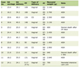 Click to view | Table 1. Multivariate Analysis for CAVI |
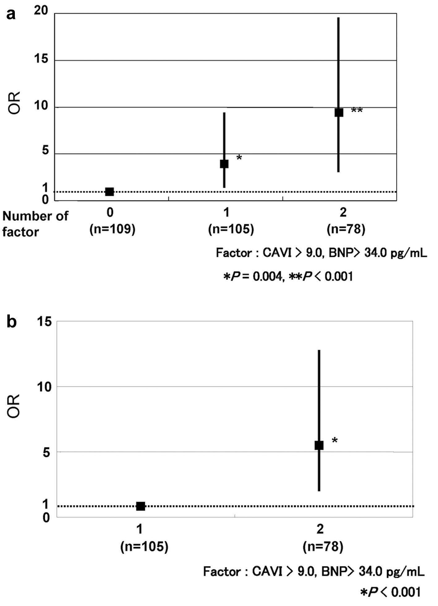 Click for large image | Figure 1. Association between the combination of high CAVI and high BNP and the presence of stage B heart failure. (a) Adjusted odds ratios (ORs) and 95% confidence intervals (CIs) for stage B heart failure according to the number of risk factors (0, 1, or 2), with the 0-factor group as reference. Compared to patients with low CAVI and low BNP (0 factors), those with one high parameter (one factor) showed a 3.81-fold higher odds (OR: 3.81; 95% CI: 1.54–9.41, *P = 0.004), and those with both high parameters (two factors) showed a 9.72-fold higher odds (OR: 9.72; 95% CI: 3.24–19.92, P < 0.001). (b) Comparison of the adjusted OR between subjects with both risk factors (two factors) and those with only one risk factor (one factor) as the reference. The adjusted OR for the two-factor group was 5.36 (95% CI: 2.39–12.90, P < 0.001). Risk factors are defined as CAVI > 9.0 and BNP > 34.0 pg/mL. Adjustment factor: calf circumference, RAS inhibitors, E/e' ratio, skin autofluorescence, SEVR, GLP-1 receptor agonists, age, eGFR, CAD, SGLT2 inhibitors. Data expressed OR and 95% CI. HF: heart failure; CAVI: cardio-ankle vascular index; BNP: brain natriuretic peptide; OR: odds ratio; CI: confidence interval; RAS: renin–angiotensin system; SEVR: subendocardial viability ratio; GLP-1: glucagon-like peptide-1; eGFR: estimated glomerular filtration rate; CAD: coronary artery disease; SGLT2: sodium glucose cotransporter 2. |
| Results | ▴Top |
Baseline clinical characteristics
Table 2 indicates the baseline clinical characteristics of the study participants. The CAVI ranged from 5.2 to 14.1, with an average value of 9.2. Regarding the HF stage, 157 patients (54%) were in stage A HF, and 135 patients (46%) were in stage B HF. The average HbA1c level was 7.1%. In total, 175 patients (60%) were taking RAS inhibitors. However, regarding anti-diabetes medications, 214 patients (73%) were taking dipeptidyl peptidase 4 inhibitors, while SGLT2 inhibitors use and GLP-1 receptor agonists use were found in 78 patients (27%) and 23 patients (8%), respectively.
Click to view | Table 2. Baseline Characteristics of the Study Population |
Correlations between CAVI and various clinical parameters
Table 3 presents the correlations between CAVI and various clinical parameters. CAVI had significantly positive correlations with age, stage B HF, current smoking, history of CAD, interventricular septal thickness at end-diastole, left atrial dimension, E/e' ratio, hemoglobin A1c, skin autofluorescence, BNP, and hs-CRP. However, it had significantly negative correlations with obesity, eGFR, SEVR, calf circumference, RAS inhibitors use, SGLT2 inhibitors use, and GLP-1 receptor agonists use.
Click to view | Table 3. Correlation Between CAVI and Various Clinical Characteristics |
Multiple regression analysis
Table 1 presents the results of the multivariable regression analysis for CAVI. Of the 18 explanatory variables associated with CAVI by simple correlation, 11 explanatory factors (calf circumference, RAS inhibitors use, E/e' ratio, skin autofluorescence, SEVR, GLP-1 receptor agonists use, age, eGFR, CAD, stage B HF, and SGLT2 inhibitors use) had independent associations with CAVI (R2 = 0.663). However, BNP was not selected as independent variables for CAVI as dependent variable.
CAVI threshold of the combination of CAVI and BNP for distinguishing between stage A HF and stage B HF
Figure 2 presents CAVI and BNP thresholds for distinguishing between stage A HF and stage B HF via the ROC curve analysis using the Youden index. This analysis indicated that 9.0 is the CAVI threshold for distinguishing between the two HF stages (Fig. 2a). For BNP, 34.0 pg/mL was the threshold for distinguishing between the two HF stages (Fig. 2b). Figure 1 indicates the combination of CAVI and BNP to distinguish stage A HF and stage B HF using multiple logistic regression analysis. Compared to patients with low CAVI and low BNP, patients for whom one of these parameters was high showed a 3.81-fold higher odds (OR: 3.81; 95% confidence interval (CI): 1.54–9.41, P = 0.004), and those in whom both parameters were high showed 9.72-fold higher odds (OR: 9.72; 95% confidence interval: 3.24–19.92, P < 0.001, Fig. 1a). In the comparison of the adjusted OR between subjects with both risk factors (two factors) and those with only one risk factor (one factor) as the reference, the adjusted OR for the two-factor group was 5.36 (95% CI: 2.39–12.90, P < 0.001, Fig. 1b).
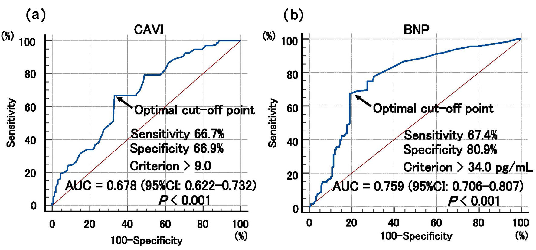 Click for large image | Figure 2. The optimal cut-off values of CAVI and BNP threshold for distinguishing between stage A HF and stage B HF using the receiver operating characteristic curve analysis. This analysis indicated that 9.0 is the CAVI threshold for distinguishing between the two HF stages (a). For BNP, 34.0 pg/mL was the threshold for distinguishing between the two HF stages (b). Comparison between the AUC of CAVI and BNP was performed using the DeLong test (P = 0.073). CAVI: cardio-ankle vascular index; BNP: brain natriuretic peptide; HF: heart failure; AUC: area under the curve; CI: confidence interval. |
| Discussion | ▴Top |
This study examined the clinical significance of CAVI, an indicator of arterial stiffness, in stage A/B HF patients with T2DM. Although CAVI was associated with various clinical parameters, the multiple regression analysis revealed independent associations with 11 factors by high levels of the multiple correlation coefficient, as R2 = 0.663. Of these, the associations between age and CAD and CAVI were consistent with previous reports [23–25]. Regarding the relationship between cardiac function indicators and CAVI, independent associations were found with E/e' (an indicator of diastolic function) and SEVR (a representation of subendocardial ischemia). The importance of AGEs was demonstrated in the association between blood glucose-related factors and CAVI. Furthermore, an independent association was found with decrease in eGFR (an indicator of renal function), suggesting that CAVI is an important factor connecting the cardio-renal interaction in stage A/B HF patients with T2DM. In addition, calf circumference, an indicator of sarcopenia, was selected as the strongest explanatory factor for CAVI in this study. Regarding medications, independent associations were found between CAVI and novel anti-diabetes medications, such as SGLT2 inhibitors and GLP-1 receptor agonists, in addition to RAS inhibitors. However, another important finding obtained in this study was the usefulness of evaluating CAVI and BNP together in distinguishing between stage A HF and stage B HF in patients with T2DM.
Relationships among left ventricular diastolic dysfunction, subendocardial ischemia, and CAVI
In recent years, the importance of left ventricular diastolic function in cases with preserved left ventricular systolic function has been demonstrated in HF pathogenesis. However, the mean LVEF in the study group was 62.6%, suggesting that most cases had preserved left ventricular systolic function, highlighting the importance of left ventricular diastolic function in this study group. In addition, there have been several reports showing a correlation between elevated arterial stiffness and left ventricular diastolic function, and several reports have shown a correlation between CAVI and markers of diastolic function, including the E/e' ratio [26, 27], indicating that the importance of increase in CAVI as a marker of arterial stiffness for diastolic dysfunction. This may be explained by the ventricular-vascular coupling mechanism, which demonstrates how increased arterial stiffness (high CAVI) elevates left ventricular afterload and alters myocardial properties, which ultimately leads to left ventricular diastolic dysfunction and a subsequent increase in left ventricular filling pressure (high E/e'). In addition, the results of this study support the validity of existing reports, and the present findings suggest that monitoring CAVI from an early stage may help identify patients with subclinical diastolic dysfunction. However, due to the cross-sectional design of this study, it remains unclear whether maintaining a lower CAVI can directly prevent the progression of diastolic dysfunction in stage A/B HF patients with T2DM. As mentioned above, SEVR is a finding indicating subendocardial ischemia, and, in recent years, there have been reports that decreased SEVR is associated with HF progression [28, 29]. Furthermore, Han et al demonstrated that arterial stiffness is associated with decreased SEVR [30]. This study also demonstrated an independent involvement of CAVI elevation in the decrease in SEVR in stage A/B HF patients with T2DM, suggesting that preventing an increase in CAVI is important from the perspective of subendocardial ischemia. However, due to its cross-sectional design, this study could not determine the incidence of HF; therefore, further prospective studies are required to evaluate reductions in HF incidence by increased SEVR through CAVI improvement in patients with T2DM.
AGEs, arterial stiffness, and HF
AGEs are deeply involved in various pathophysiological aspects in systemic organs of patients with T2DM, and several studies have indicated that AGEs and AGEs receptors are involved in HF pathogenesis [31–33]. In addition, existing reports have indicated that skin autofluorescence is involved in HF pathogenesis [34–36]. However, a significant association has been reported between skin autofluorescence and arterial stiffness including CAVI [37, 38], suggesting that AGEs act on vascular stiffness through the pathway of inflammation and oxidative stress in vascular wall. In this study as well, among the blood glucose-related factors, only skin autofluorescence was identified as an independent determinant of CAVI, an indicator of arterial stiffness in stage A/B HF patients with T2DM. This indicates a close association between AGEs and arterial stiffness, as reflected in CAVI, in stage A/B HF patients with T2DM. Although these findings do not prove a causal relationship or that treating AGEs will prevent the development of symptomatic HF, they highlight a potential pathophysiological link that warrants further prospective investigation.
Sarcopenia and CAVI
Several basic and clinical studies have highlighted the importance of sarcopenia in HF pathogenesis, with inflammation, oxidative stress, and sympathetic nervous system activation being cited as contributing factors [39, 40]. In addition, several researchers have reported an association between sarcopenia and vascular function, inferring the pathogenesis of HF from a decline in vascular function [41, 42]. Meanwhile, several previous studies have shown an association between CAVI and sarcopenia [43–45], supporting the existence of a close relationship between the two. In this study as well, calf circumference, an indicator of sarcopenia, was selected as the strongest contributing factor to CAVI, even though reliability of calf circumference as a marker of sarcopenia. Therefore, considering existing reports alongside the results of this study, the findings demonstrate a significant statistical association between sarcopenia and vascular dysfunction (including an increase in CAVI) as interrelated features in stage A/B HF patients with T2DM. This association may be mediated by shared biological mechanisms, such as chronic inflammation, oxidative stress, and altered adipokine profiles. While active detection of sarcopenia may be useful for comprehensive risk stratification, it should be noted that calf circumference primarily reflects muscle quantity. A definitive diagnosis of sarcopenia requires a comprehensive assessment of muscle quality, including muscle strength and physical function. Furthermore, longitudinal studies are necessary to elucidate causal direction and to confirm whether clinical interventions for sarcopenia can actually reduce the future incidence of HF in this population.
Medications and CAVI
RAS inhibitors are known to be involved in various pathological conditions associated with T2DM, and their use is recommended for patients with this metabolic disease. However, several researchers have reported that RAS inhibitors reduce CAVI [46, 47], and this study’s findings are consistent with existing reports. However, the effect of RAS inhibitors on HF with preserved ejection fraction is limited [48, 49]. Since the study group mainly consisted of cases with preserved LVEF, it is likely that the effect of RAS inhibitors on HF prevention is limited. Nevertheless, it is expected that RAS inhibitor use in patients with high CAVI may efficiently lead to the development of HF. To confirm this in the future, the usefulness of RAS inhibitors in preventing HF in cases with preserved LVEF should be investigated from the perspective of CAVI. Recently, SGLT2 inhibitors and GLP-1 receptor agonists have attracted attention as novel T2DM treatment options. Both medications are attracting attention for their additional benefit of acting on the cardiovascular system [50, 51], and an independent relationship between both medications and CAVI was observed in this cross-sectional study. Sakai et al reported that SGLT2 inhibitors such as empagliflozin, luseogliflozin, and tofogliflozin lower CAVI [52]. However, to the best of the author’s knowledge, there are no reports showing an association between GLP-1 receptor agonists and CAVI. Nevertheless, a negative correlation between CAVI and GLP-1 receptor agonists was observed in the multivariate analysis of this study. Several reports describe the mechanisms by which GLP-1 receptor agonists affect vascular function such as vascular fibrosis, endothelial dysfunction, inflammation [53, 54], and oxidative stress, and these underlying pathways might support a potential link with arterial stiffness. In addition, despite its cross-sectional design, the results of this study suggest a potential association between GLP-1 receptor agonist use and lower CAVI. Hence, as detailed in the “Limitations” section, prospective interventional studies are mandatory to confirm whether this reflects a true medication effect. Furthermore, because recent clinical evidence suggests that SGLT2 inhibitors and GLP-1 receptor agonists may potentially reduce skeletal muscle mass or lean body mass, clinicians should be cautious regarding the risk of sarcopenia progression [55, 56]. Consequently, the implementation of appropriate protein intake and structured exercise regimens is highly recommended for patients with T2DM when prescribing these two classes of medications.
Combination of CAVI and BNP to distinguish stage A HF and stage B HF
In this study, a significant positive correlation was observed between CAVI and BNP in a simple correlation analysis, although this association was not statistically significant in the multivariate analysis. This suggests that CAVI reflects pathological conditions involved in HF progression that cannot be captured by BNP alone in early-stage HF. Therefore, the author first determined cut-off values for distinguishing between stage A HF and stage B HF from the ROC curve analysis. As a result, the author calculated cut-off values of 9.0 for CAVI and 34 pg/mL for BNP. A CAVI of 9 was consistent with existing reports that, although the target groups differ, CAVI is generally considered a risk factor for CVD [16, 57, 58]. However, several recent reports have used BNP values of ≥ 35 pg/mL to distinguish between stage A HF and stage B HF, which is similar to the value in this study [59, 60]. Therefore, the findings support the validity of the current guideline cutoff values for BNP levels in patients with T2DM in routine clinical practice. Next, using these thresholds, the author performed a multiple logistic regression analysis. As the number of factors (high CAVI, high BNP) increased, the OR increased compared with lower levels of both parameters. Needless to say, numerous factors are involved in the progression of HF, and it is difficult to completely distinguish between stage A and stage B HF using only two markers as CAVI and BNP. However, the results of this study suggest that there are cases in which progression from stage A to stage B HF can be prevented by combining CAVI and BNP. Although the multiple regression in Figure 1 carried a risk of over-adjustment, a simplified sensitivity analysis adjusting only for core demographic factors yielded consistent and robust ORs. While the prospective multicenter study by Miyoshi et al [18] (previously cited in the “Introduction”) demonstrated the long-term prognostic value of CAVI for future cardiovascular outcomes, the present study uniquely adds to the literature by focusing strictly on a highly selected population of patients with T2DM. Furthermore, this study demonstrates the immediate clinical utility of combining CAVI with BNP levels to specifically stratify the risk of subclinical progression from stage A to stage B HF. This combined approach provides primary care clinicians with an actionable screening strategy within routine diabetic management, complementary to the broader prognostic framework established by Miyoshi et al. A notable methodological strength of the present study lies in the group assignment criteria for stage A and B HF. The author intentionally relied strictly on structural and clinical parameters—rather than BNP levels—for the initial classification. By doing so, the author successfully avoided circular reasoning when evaluating the subsequent combined utility of CAVI and BNP. Consequently, the remarkably high OR (9.72-fold) observed in patients with both high CAVI and high BNP represents a clinically objective and robust finding, free from selection bias.
Limitations
This study has several noteworthy limitations. First, it was conducted only on East Asian stage A/B HF patients with T2DM, focusing on a relatively small number of cases at a single facility. Therefore, future larger-scale, multicenter studies are anticipated to include other ethnicities to produce more generalizable findings. Second, the observed association between medication use, particularly GLP-1 receptor agonists, and lower CAVI values must be interpreted with caution. These findings are subject to confounding by indication inherent in non-randomized, cross-sectional designs. Given that GLP-1 receptor agonists were used by only 23 participants (8% of the cohort), this specific finding should be considered purely hypothesis-generating and requires validation in large-scale randomized controlled trial. Future large-scale, multicenter studies with a larger sample size of GLP-1 receptor agonists users are needed to confirm these findings. Third, the diagnostic accuracy of CAVI as a standalone screening tool for distinguishing between stage A and stage B HF was modest, as demonstrated by an AUC of 0.678, with a sensitivity of 66.7% and a specificity of 66.9%. Therefore, the threshold of ≤ 9.0 identified in the present study must be interpreted strictly as an exploratory cut-point requiring future external validation, rather than an established therapeutic target. Additionally, it should be noted that the ROC-derived optimal BNP cut-off value of 34.0 pg/mL sits slightly above the median BNP level of 29.0 pg/mL for the entire study population. This baseline distribution may limit the immediate generalizability of this specific threshold to other cohorts with different background demographics, although it represents a clinically practical screening value for the present study population. Fourth, due to the relatively small sample size and cross-sectional design of this single-center study, the potential for residual confounding from other unmeasured disease processes cannot be fully eliminated. Consequently, while the independent statistical association of CAVI and BNP suggests they may reflect distinct pathophysiological pathways (macrovascular stiffness versus myocardial wall stress), concluding that their combination provides superior clinical risk stratification remains a hypothesis that requires validation. Prospective, large-scale longitudinal studies are mandatory to confirm the clinical utility of this dual-marker approach in routine practice. Finally, while this study yielded some important insights into the significance of CAVI in stage A/B HF patients with T2DM, some questions remain. Therefore, further detailed research from both clinical and basic research perspectives is needed to clarify the significance of CAVI in stage A/B HF patients with T2DM. In addition, the validity of the results of this study needs to be verified through prospective trials. However, this study focused solely on the association between CAVI and stage A/B HF. Therefore, further research is needed regarding the association between CAVI and other diabetic complications, particularly those in the early stages.
Conclusions
This study suggests that CAVI is closely associated with pathological indicators in stage A/B HF patients with T2DM. Furthermore, the findings indicate that a combination of CAVI and BNP measurements could serve as a potential screening tool for risk stratification in the pre-symptomatic stage. Given the cross-sectional nature of this study, caution is warranted in inferring causality, and the author cannot conclude that therapeutic interventions targeting CAVI directly prevent HF progression; nevertheless, this combination holds potential for early risk stratification. To confirm these findings and improve generalizability, future large-scale, prospective multicenter clinical trials are warranted.
Acknowledgments
None to declare.
Financial Disclosure
None to declare.
Conflict of Interest
None to declare.
Informed Consent
Informed consent was obtained from all study participants.
Author Contributions
Takashi Hitsumoto contributed to research planning, data acquisition and analysis, and manuscript writing and editing.
Data Availability
The author declares that data supporting the findings of this study are available within the article.
Abbreviations
AGEs: advanced glycation end products; AU: arbitrary unit; AUC: area under the curve; BNP: brain natriuretic peptide; CAVI: cardio-ankle vascular index; CAD: coronary artery disease; CVD: ; cardiovascular disease: ; eGFR: estimated glomerular filtration rate; GLP-1: glucagon-like peptide-1; HF: heart failure; LVEF: left ventricular ejection fraction; OR: odds ratio; RAS: renin–angiotensin system; SEVR: subendocardial viability ratio; SGLT2: sodium glucose cotransporter 2; T2DM: type 2 diabetes mellitus
| References | ▴Top |
- Shimokawa H, Miura M, Nochioka K, Sakata Y. Heart failure as a general pandemic in Asia. Eur J Heart Fail. 2015;17(9):884-892.
doi pubmed - Savarese G, Becher PM, Lund LH, Seferovic P, Rosano GMC, Coats AJS. Global burden of heart failure: a comprehensive and updated review of epidemiology. Cardiovasc Res. 2023;118(17):3272-3287.
doi pubmed - Krumholz HM, Parent EM, Tu N, Vaccarino V, Wang Y, Radford MJ, Hennen J. Readmission after hospitalization for congestive heart failure among Medicare beneficiaries. Arch Intern Med. 1997;157(1):99-104.
pubmed - Palazzuoli A, Iacoviello M. Diabetes leading to heart failure and heart failure leading to diabetes: epidemiological and clinical evidence. Heart Fail Rev. 2023;28(3):585-596.
doi pubmed - Kong MG, Jang SY, Jang J, Cho HJ, Lee S, Lee SE, Kim KH, et al. Impact of diabetes mellitus on mortality in patients with acute heart failure: a prospective cohort study. Cardiovasc Diabetol. 2020;19(1):49.
doi pubmed - Mueller C, McDonald K, de Boer RA, Maisel A, Cleland JGF, Kozhuharov N, Coats AJS, et al. Heart Failure Association of the European Society of Cardiology practical guidance on the use of natriuretic peptide concentrations. Eur J Heart Fail. 2019;21(6):715-731.
doi pubmed - Tsutsui H, Albert NM, Coats AJS, Anker SD, Bayes-Genis A, Butler J, Chioncel O, et al. Natriuretic peptides: role in the diagnosis and management of heart failure: a scientific statement from the Heart Failure Association of the European Society of Cardiology, Heart Failure Society of America and Japanese Heart Failure Society. J Card Fail. 2023;29(5):787-804.
doi pubmed - Hitsumoto T. Efficacy of the reactive oxygen metabolite test as a predictor of initial heart failure hospitalization in elderly patients with chronic heart failure. Cardiol Res. 2018;9(3):153-160.
doi pubmed - Shirai K, Utino J, Otsuka K, Takata M. A novel blood pressure-independent arterial wall stiffness parameter; cardio-ankle vascular index (CAVI). J Atheroscler Thromb. 2006;13(2):101-107.
doi pubmed - Hitsumoto T. Clinical significance of cardio-ankle vascular index as a cardiovascular risk factor in elderly patients with type 2 diabetes mellitus. J Clin Med Res. 2018;10(4):330-336.
doi pubmed - Kim KJ, Lee BW, Kim HM, Shin JY, Kang ES, Cha BS, Lee EJ, et al. Associations between cardio-ankle vascular index and microvascular complications in type 2 diabetes mellitus patients. J Atheroscler Thromb. 2011;18(4):328-336.
doi pubmed - Yamaguchi T, Shirai K, Nagayama D, Nakamura S, Oka R, Tanaka S, Watanabe Y, et al. Bezafibrate ameliorates arterial stiffness assessed by cardio-ankle vascular index in hypertriglyceridemic patients with type 2 diabetes mellitus. J Atheroscler Thromb. 2019;26(7):659-669.
doi pubmed - Sato Y, Nagayama D, Saiki A, Watanabe R, Watanabe Y, Imamura H, Yamaguchi T, et al. Cardio-ankle vascular index is independently associated with future cardiovascular events in outpatients with metabolic disorders. J Atheroscler Thromb. 2016;23(5):596-605.
doi pubmed - Nakamura K, Tomaru T, Yamamura S, Miyashita Y, Shirai K, Noike H. Cardio-ankle vascular index is a candidate predictor of coronary atherosclerosis. Circ J. 2008;72(4):598-604.
doi pubmed - Miyoshi T, Ito H, Shirai K, Horinaka S, Higaki J, Yamamura S, Saiki A, et al. Predictive value of the cardio-ankle vascular index for cardiovascular events in patients at cardiovascular risk. J Am Heart Assoc. 2021;10(16):e020103.
doi pubmed - Hitsumoto T. Clinical significance of the cardio-ankle vascular index as a cardiovascular disease risk factor in Japanese elderly patients with obesity. J Clin Med Res. 2025;17(9):518-528.
doi pubmed - Schillaci G, Battista F, Settimi L, Anastasio F, Pucci G. Cardio-ankle vascular index and subclinical heart disease. Hypertens Res. 2015;38(1):68-73.
doi pubmed - Miyoshi T, Shirai K, Horinaka S, Higaki J, Yamamura S, Saiki A, Takahashi M, et al. Association between cardio-ankle vascular index and heart failure outcomes: insights from a prospective multicenter cohort. JACC Adv. 2025;4(12 Pt 2):102187.
doi pubmed - Yancy CW, Jessup M, Bozkurt B, Butler J, Casey DE, Jr., Drazner MH, Fonarow GC, et al. 2013 ACCF/AHA guideline for the management of heart failure: a report of the American College of Cardiology Foundation/American Heart Association Task Force on Practice Guidelines. J Am Coll Cardiol. 2013;62(16):e147-239.
doi pubmed - Saiki A, Sato Y, Watanabe R, Watanabe Y, Imamura H, Yamaguchi T, Ban N, et al. The role of a novel arterial stiffness parameter, Cardio-Ankle Vascular Index (CAVI), as a surrogate marker for cardiovascular diseases. J Atheroscler Thromb. 2016;23(2):155-168.
doi pubmed - Imai E, Horio M, Nitta K, Yamagata K, Iseki K, Hara S, Ura N, et al. Estimation of glomerular filtration rate by the MDRD study equation modified for Japanese patients with chronic kidney disease. Clin Exp Nephrol. 2007;11(1):41-50.
doi pubmed - Schisterman EF, Perkins NJ, Liu A, Bondell H. Optimal cut-point and its corresponding Youden Index to discriminate individuals using pooled blood samples. Epidemiology. 2005;16(1):73-81.
doi pubmed - Shirai K, Hiruta N, Song M, Kurosu T, Suzuki J, Tomaru T, Miyashita Y, et al. Cardio-ankle vascular index (CAVI) as a novel indicator of arterial stiffness: theory, evidence and perspectives. J Atheroscler Thromb. 2011;18(11):924-938.
doi pubmed - Mineoka Y, Fukui M, Tanaka M, Tomiyasu K, Akabame S, Nakano K, Yamazaki M, et al. Relationship between cardio-ankle vascular index (CAVI) and coronary artery calcification (CAC) in patients with type 2 diabetes mellitus. Heart Vessels. 2012;27(2):160-165.
doi pubmed - Dung LV, Son PN, Nguyen KLT, Pho DC, Thang NM, Thang DC, Son PT. Relationship between Cardio-Ankle Vascular Index (CAVI) and the severity of coronary artery lesions: a case-control study. Vasc Health Risk Manag. 2025;21:607-615.
doi pubmed - Kim H, Kim HS, Yoon HJ, Park HS, Cho YK, Nam CW, Hur SH, et al. Association of cardio-ankle vascular index with diastolic heart function in hypertensive patients. Clin Exp Hypertens. 2014;36(4):200-205.
doi pubmed - Nguyen TH, Nakamura T, Horikoshi T, Uematsu M, Kobayashi T, Yoshizaki T, Watanabe Y, et al. Arterial stiffness and left ventricular diastolic dysfunction: insights from the cardio-ankle vascular index. Heart Lung. 2026;78:102741.
doi pubmed - Masutani S, Kuwata S, Kurishima C, Iwamoto Y, Saiki H, Sugimoto M, Ishido H, et al. Ventricular-vascular dynamics in pediatric patients with heart failure and preserved ejection fraction. Int J Cardiol. 2016;225:306-312.
doi pubmed - Hommo H, Sugawara T, Ueno H, Kawashima H, Uchida K, Minegishi S, Chen L, et al. Association of arterial velocity pulse index and arterial pressure-volume index with central arterial stiffness and cardiac function in the Japanese population. J Clin Med. 2026;15(4).
doi pubmed - Han J, Tang J, Zhao Y, Xiong J, Zhao S, Yue W, Xu Y, et al. Subendocardial viability ratio is associated with target organ damage and hints at a potential independent predictor of cardiovascular mortality in older adults: a prospective cohort study. J Am Heart Assoc. 2026;15(1):e043643.
doi pubmed - Ma H, Li SY, Xu P, Babcock SA, Dolence EK, Brownlee M, Li J, et al. Advanced glycation endproduct (AGE) accumulation and AGE receptor (RAGE) up-regulation contribute to the onset of diabetic cardiomyopathy. J Cell Mol Med. 2009;13(8B):1751-1764.
doi pubmed - Hitsumoto T. Clinical significance of skin autofluorescence in elderly patients with long-standing persistent atrial fibrillation. Cardiol Res. 2019;10(3):181-187.
doi pubmed - Bucciarelli LG, Ananthakrishnan R, Hwang YC, Kaneko M, Song F, Sell DR, Strauch C, et al. RAGE and modulation of ischemic injury in the diabetic myocardium. Diabetes. 2008;57(7):1941-1951.
doi pubmed - Hitsumoto T. Clinical significance of skin autofluorescence in patients with type 2 diabetes mellitus with chronic heart failure. Cardiol Res. 2018;9(2):83-89.
doi pubmed - Yoshioka K. Skin autofluorescence is associated with high-sensitive cardiac troponin T, a circulating cardiac biomarker, in Japanese patients with diabetes: A cross-sectional study. Diab Vasc Dis Res. 2018;15(6):559-566.
doi pubmed - Hitsumoto T. Skin autofluorescence as a predictor of first heart failure hospitalization in patients with heart failure with preserved ejection fraction. Cardiol Res. 2020;11(4):247-255.
doi pubmed - Birukov A, Cuadrat R, Polemiti E, Eichelmann F, Schulze MB. Advanced glycation end-products, measured as skin autofluorescence, associate with vascular stiffness in diabetic, pre-diabetic and normoglycemic individuals: a cross-sectional study. Cardiovasc Diabetol. 2021;20(1):110.
doi pubmed - van Eupen MG, Schram MT, van Sloten TT, Scheijen J, Sep SJ, van der Kallen CJ, Dagnelie PC, et al. Skin autofluorescence and pentosidine are associated with aortic stiffening: the Maastricht study. Hypertension. 2016;68(4):956-963.
doi pubmed - Collamati A, Marzetti E, Calvani R, Tosato M, D'Angelo E, Sisto AN, Landi F. Sarcopenia in heart failure: mechanisms and therapeutic strategies. J Geriatr Cardiol. 2016;13(7):615-624.
doi pubmed - Curcio F, Testa G, Liguori I, Papillo M, Flocco V, Panicara V, Galizia G, et al. Sarcopenia and heart failure. Nutrients. 2020;12(1):211.
doi pubmed - Kim SR, Cho DH, Kim MN, Park SM. Rationale and study design of differences in cardiopulmonary exercise capacity according to coronary microvascular dysfunction and body composition in patients with suspected heart failure with preserved ejection fraction. Int J Heart Fail. 2021;3(4):237-243.
doi pubmed - Dos Santos MR, Saitoh M, Ebner N, Valentova M, Konishi M, Ishida J, Emami A, et al. Sarcopenia and endothelial function in patients with chronic heart failure: results from the studies investigating comorbidities aggravating heart failure (SICA-HF). J Am Med Dir Assoc. 2017;18(3):240-245.
doi pubmed - Kirkham FA, Bunting E, Fantin F, Zamboni M, Rajkumar C. Independent Association Between Cardio-Ankle Vascular Index and Sarcopenia in Older U.K. Adults. J Am Geriatr Soc. 2019;67(2):317-322.
doi pubmed - Park HE, Chung GE, Lee H, Kim MJ, Choi SY, Lee W, Yoon JW. Significance of low muscle mass on arterial stiffness as measured by cardio-ankle vascular index. Front Cardiovasc Med. 2022;9:857871.
doi pubmed - Ogawa A, Shimizu K, Nakagami T, Maruoka H, Shirai K. Physical function and cardio-ankle vascular index in elderly heart failure patients. Int Heart J. 2020;61(4):769-775.
doi pubmed - Miyashita Y, Saiki A, Endo K, Ban N, Yamaguchi T, Kawana H, Nagayama D, et al. Effects of olmesartan, an angiotensin II receptor blocker, and amlodipine, a calcium channel blocker, on Cardio-Ankle Vascular Index (CAVI) in type 2 diabetic patients with hypertension. J Atheroscler Thromb. 2009;16(5):621-626.
doi pubmed - Kinouchi K, Ichihara A, Sakoda M, Kurauchi-Mito A, Murohashi-Bokuda K, Itoh H. Effects of telmisartan on arterial stiffness assessed by the cardio-ankle vascular index in hypertensive patients. Kidney Blood Press Res. 2010;33(4):304-312.
doi pubmed - Yusuf S, Pfeffer MA, Swedberg K, Granger CB, Held P, McMurray JJ, Michelson EL, et al. Effects of candesartan in patients with chronic heart failure and preserved left-ventricular ejection fraction: the CHARM-Preserved Trial. Lancet. 2003;362(9386):777-781.
doi pubmed - Massie BM, Carson PE, McMurray JJ, Komajda M, McKelvie R, Zile MR, Anderson S, et al. Irbesartan in patients with heart failure and preserved ejection fraction. N Engl J Med. 2008;359(23):2456-2467.
doi pubmed - Anker SD, Butler J, Filippatos G, Ferreira JP, Bocchi E, Bohm M, Brunner-La Rocca HP, et al. Empagliflozin in heart failure with a preserved ejection fraction. N Engl J Med. 2021;385(16):1451-1461.
doi pubmed - Marso SP, Daniels GH, Brown-Frandsen K, Kristensen P, Mann JF, Nauck MA, Nissen SE, et al. Liraglutide and cardiovascular outcomes in type 2 diabetes. N Engl J Med. 2016;375(4):311-322.
doi pubmed - Sakai T, Miura S. Effects of sodium-glucose cotransporter 2 inhibitor on vascular endothelial and diastolic function in heart failure with preserved ejection fraction - novel prospective cohort study. Circ Rep. 2019;1(7):286-295.
doi pubmed - Helmstadter J, Frenis K, Filippou K, Grill A, Dib M, Kalinovic S, Pawelke F, et al. Endothelial GLP-1 (Glucagon-Like Peptide-1) receptor mediates cardiovascular protection by liraglutide in mice with experimental arterial hypertension. Arterioscler Thromb Vasc Biol. 2020;40(1):145-158.
doi pubmed - Battistoni A, Piras L, Tartaglia N, Carrano FM, De Vitis C, Barbato E. Glucagon-like peptide-1 receptor agonists and the endothelium: molecular and clinical insights into cardiovascular protection. Front Med (Lausanne). 2025;12:1669685.
doi pubmed - Zhang S, Qi Z, Wang Y, Song D, Zhu D. Effect of sodium-glucose transporter 2 inhibitors on sarcopenia in patients with type 2 diabetes mellitus: a systematic review and meta-analysis. Front Endocrinol (Lausanne). 2023;14:1203666.
doi pubmed - Urbina J, Salinas-Ruiz LE, Valenciano C, Clapp B. Micronutrient and nutritional deficiencies associated with GLP-1 receptor agonist therapy: a narrative review. Clin Obes. 2026;16(1):e70070.
doi pubmed - Okamoto Y, Miyoshi T, Ichikawa K, Takaya Y, Nakamura K, Ito H. Cardio-ankle vascular index as an arterial stiffness marker improves the prediction of cardiovascular events in patients without cardiovascular diseases. J Cardiovasc Dev Dis. 2022;9(11):368.
doi pubmed - Miyoshi T, Ito H. Arterial stiffness in health and disease: the role of cardio-ankle vascular index. J Cardiol. 2021;78(6):493-501.
doi pubmed - Bozkurt B, Coats AJS, Tsutsui H, Abdelhamid CM, Adamopoulos S, Albert N, Anker SD, et al. Universal definition and classification of heart failure: a report of the Heart Failure Society of America, Heart Failure Association of the European Society of Cardiology, Japanese Heart Failure Society and Writing Committee of the Universal Definition of Heart Failure: Endorsed by the Canadian Heart Failure Society, Heart Failure Association of India, Cardiac Society of Australia and New Zealand, and Chinese Heart Failure Association. Eur J Heart Fail. 2021;23(3):352-380.
doi pubmed - Fujimoto W, Odajima S, Okamoto H, Iwasaki M, Nagao M, Konishi A, Shinohara M, et al. Importance of B-type natriuretic peptide in the detection of patients with structural heart disease in a primary care setting. Circ J. 2024;88(5):732-739.
doi pubmed
This article is distributed under the terms of the Creative Commons Attribution 4.0 International License (CC BY 4.0), which permits unrestricted use, distribution, and reproduction in any medium, including commercial use, provided the original work is properly cited.
Journal of Clinical Medicine Research is published by Elmer Press Inc.