Journal of Clinical Medicine Research, ISSN 1918-3003 print, 1918-3011 online, Open Access
Article copyright, the authors; Journal compilation copyright, J Clin Med Res and Elmer Press Inc
Journal website https://jocmr.elmerjournals.com

Review

Volume 16, Number 9, August 2024, pages 411-422


Potential Use of MicroRNA Technology in Thalassemia Therapy

Figures

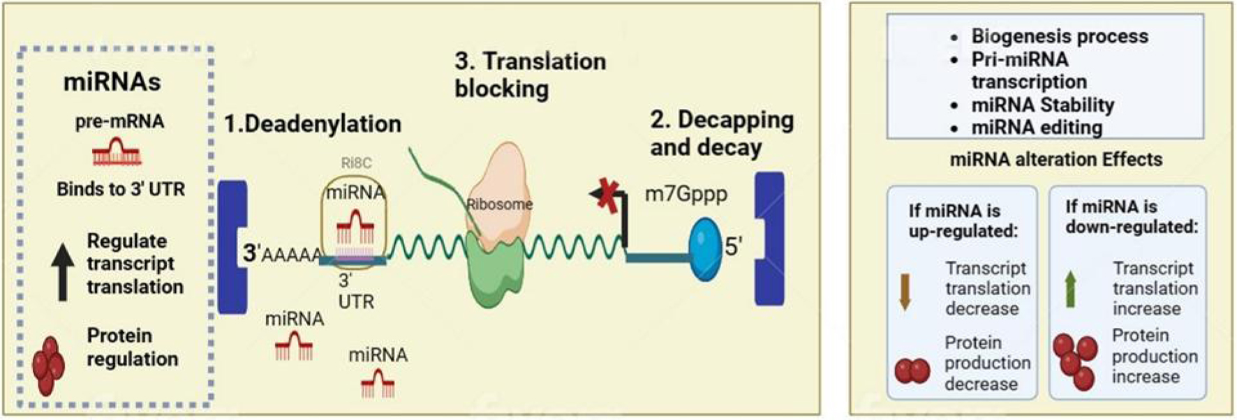
Figure 1.
Figure 1. Normal function and alteration of microRNA (miRNA) in biological mechanism. MiRNAs exert their gene-silencing effects through a sequence of molecular interactions, initiating with deadenylation, where they induce the shortening of the poly(A) tail at the mRNA's 3' end, setting the stage for mRNA degradation; they also facilitate decapping, removing the 5' cap (m7Gppp) which is essential for messenger RNA (mRNA) stability, and they can impede protein synthesis directly by blocking translation, preventing ribosomes from accessing the mRNA.
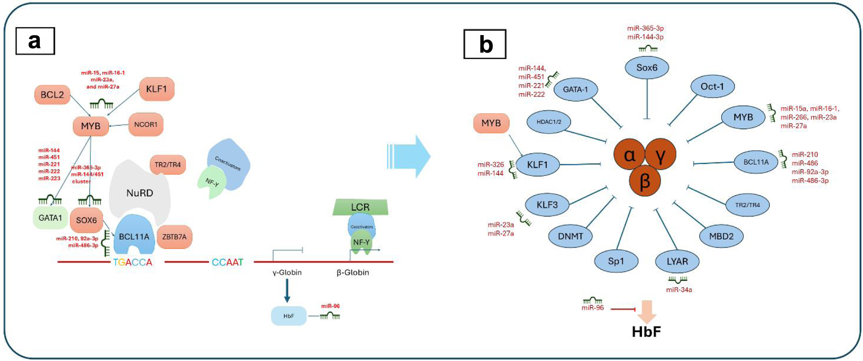
Figure 2.
Figure 2. MicroRNA (miRNA) regulation in erythropoiesis (a) and hemoglobin fetus signaling (b). (a) The complex interplay of transcription factors, miRNAs, and their targets during erythrocyte development, including key regulators such as Myb proto-oncogene protein (MYB), Kruppel-like factor 1 (KLF1), and GATA-binding factor 1 (GATA1). (b) The miRNA-mediated signaling pathways that influence fetal hemoglobin (HbF) expression, highlighting the roles of various factors like KLF1, MYB, and B-cell lymphoma/leukemia 11A (BCL11A) in modulating HbF levels through direct and indirect miRNA interactions.
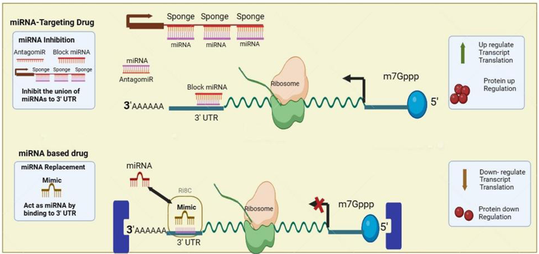
Figure 3.
Figure 3. MicroRNA (miRNA) application for thalassemia gene therapy. AntagomiRs function to inhibit miRNAs by binding and sequestering them, thus blocking their interaction with the 3' untranslated region (UTR) of messenger RNAs (mRNAs), which lifts the miRNA-mediated gene silencing and results in enhanced translation and protein production. MiRNA-based drugs utilize synthetic miRNA mimics that bind to the 3' UTR of target mRNAs to emulate natural miRNA functions, resulting in decreased mRNA translation and subsequent downregulation of protein synthesis.

Table

Table 1. List of the MiRNAs Along With Their Biological Aspects May Be Included in Thalassemia Pathophysiology
 
Name of miRNABiological aspects
miRNA: microRNA; GSH: glutathione; SOD: superoxide dismutase; NF-κB: nuclear factor kappa B; FoxO3: forkhead box protein O3; ROS: reactive oxygen species; TfR1: transferrin receptor 1.
miR-451Regulating normal erythropoiesis; downregulated in beta-thalassemia leading to erythroid hyperplasia
miR-24-1Part of miR-23b/27b/24-1 cluster suppress fibrotic of liver, disturbing hepcidin
miR-92a-3pRegulating gamma-globin, GSH, SOD, ROS, and cell apoptosis
miR-16-5pAssociated with hemolysis/transfusion reactions
miR-210Suppressing erythropoiesis in alpha-thalassemia; modulating oxidative stress
miR-let-7bControlling inflammation by regulating the NF-κB pathway
miR-20aRegulating iron metabolism by targeting ferritin; potential biomarker for iron overload and liver damage
miR-144Increased sensitivity to oxidative stress
miR-221Erythroid cell apoptosis
miR-let-7bRegulating ferroportin and iron metabolism
miR-155Modulating immune reactions associated with transfusions
miR-21Potential biomarker for iron overload and liver damage
miR-155Controlling inflammation by regulating the NF-κB pathway
miR-222Upregulated in alpha-thalassemia leading to erythroid cell apoptosis
miR-92a-3pAssociated with hemolysis/transfusion reactions
miR-27bPart of miR-23b/27b/24-1 cluster suppress fibrotic of liver
miR-9Suppressing FoxO3 and affecting ROS production
miR-485-3pRegulating iron metabolism by targeting TfR1
miR-214Upregulated in thalassemia patients with oxidative stress
miR-146aPotential biomarker for inflammation-related complications like sepsis
miR-122Regulating hepcidin and iron metabolism
miR-16Regulating normal erythropoiesis; downregulated in beta-thalassemia leading to erythroid hyperplasia
miR-150Inhibiting proliferation and differentiation of erythroid progenitor cells; modulating immune cell function (T and B lymphocytes); potential biomarker for erythropoiesis