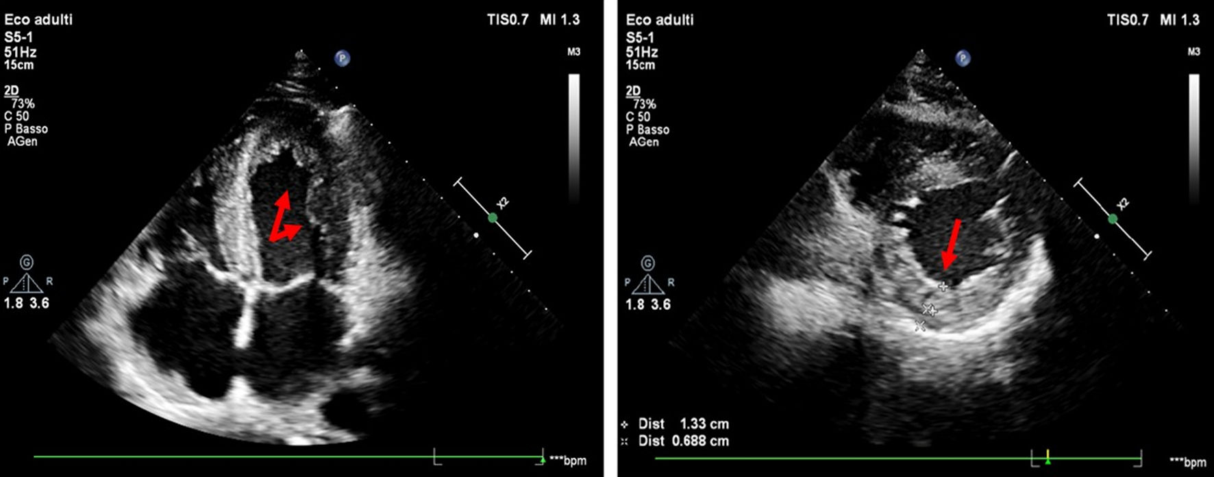
Journal of Clinical Medicine Research, ISSN 1918-3003 print, 1918-3011 online, Open Access
Article copyright, the authors; Journal compilation copyright, J Clin Med Res and Elmer Press Inc
Journal website https://jocmr.elmerjournals.com

Case Report

Volume 17, Number 1, January 2025, pages 60-65


Left Ventricular Non-Compaction, Atrial Fibrillation and ANK2 Mutation in a Young Athlete

Figures

Figure 1.
Figure 1. Echocardiographic examination (four-chamber and parasternal short-axis view) shows multiple trabeculations in the apex and lateral wall of the left ventricle, a two-layer myocardial structure with a thin compacted and thick non-compacted (NC) layer, systolic NC/C ratio > 2 (red arrows).
Figure 2.
Figure 2. Speckle tracking analysis, global longitudinal strain (-18.3%).
Figure 3.
Figure 3. The cMRI showed distinct trabeculations localized to the lateral wall, and part of the posterior wall, as well as the apex (red arrows). cMRI: cardiac magnetic resonance imaging.
Figure 4.
Figure 4. Raw data visualization of the variant c.9145C>T, p.Arg3049Trp in the ANK2 gene (NM_001148) on IGV (Integrative Genomics Viewer). ANK2: ankyrin 2; IGV: Integrative Genomics Viewer.