Journal of Clinical Medicine Research, ISSN 1918-3003 print, 1918-3011 online, Open Access
Article copyright, the authors; Journal compilation copyright, J Clin Med Res and Elmer Press Inc
Journal website https://jocmr.elmerjournals.com

Review

Volume 17, Number 7, July 2025, pages 365-374


Office-Based Middle Ear Surgery Under Local Anesthesia: A Contemporary Review

Figures

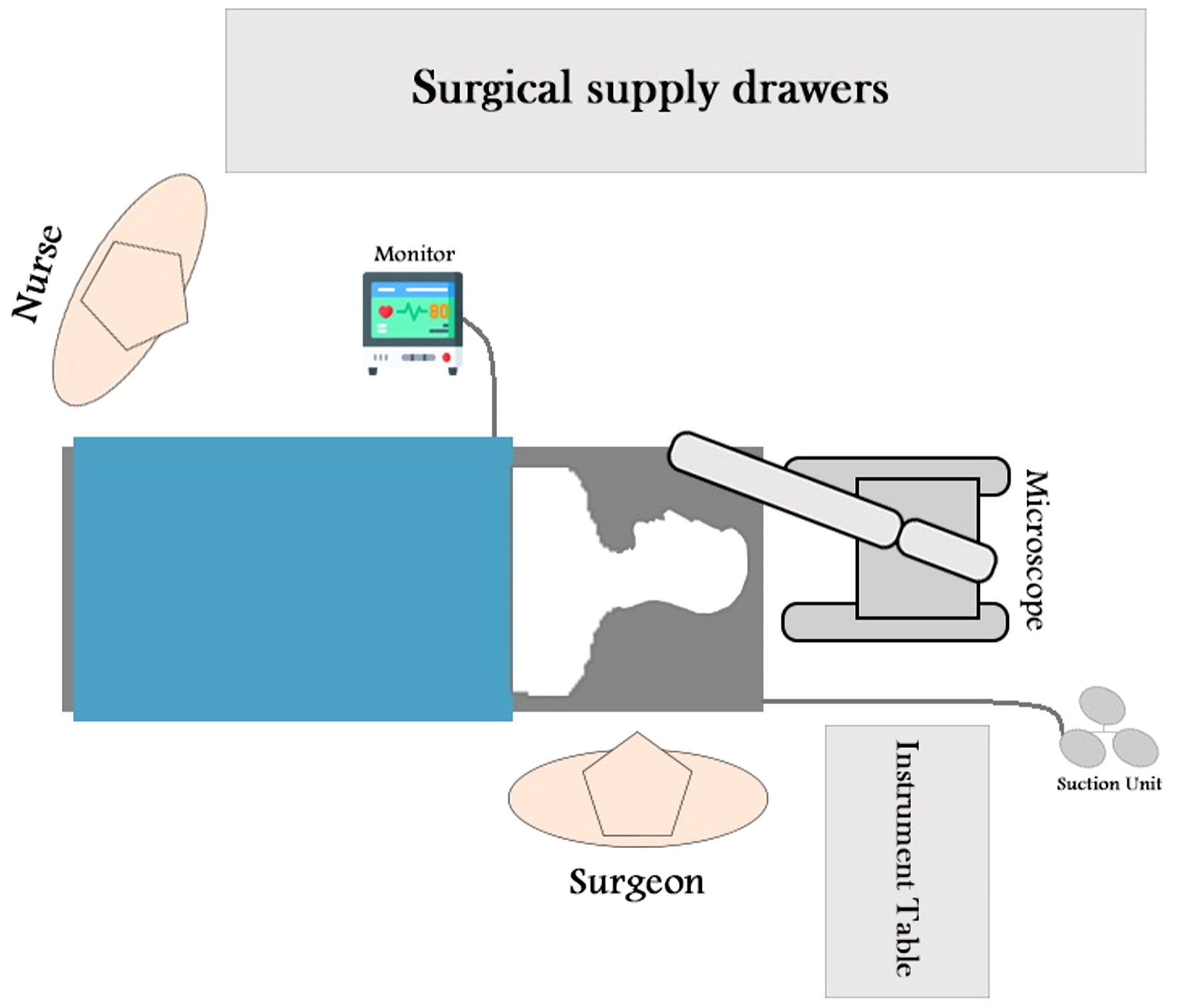
Figure 1.
Figure 1. Literature review flow diagram.
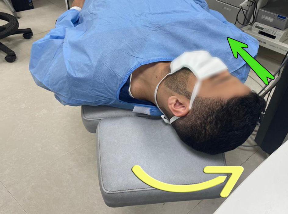
Figure 2.
Figure 2. Ergonomic configuration of an operating room, designed to enable the surgeon to perform procedures in the ear independently.
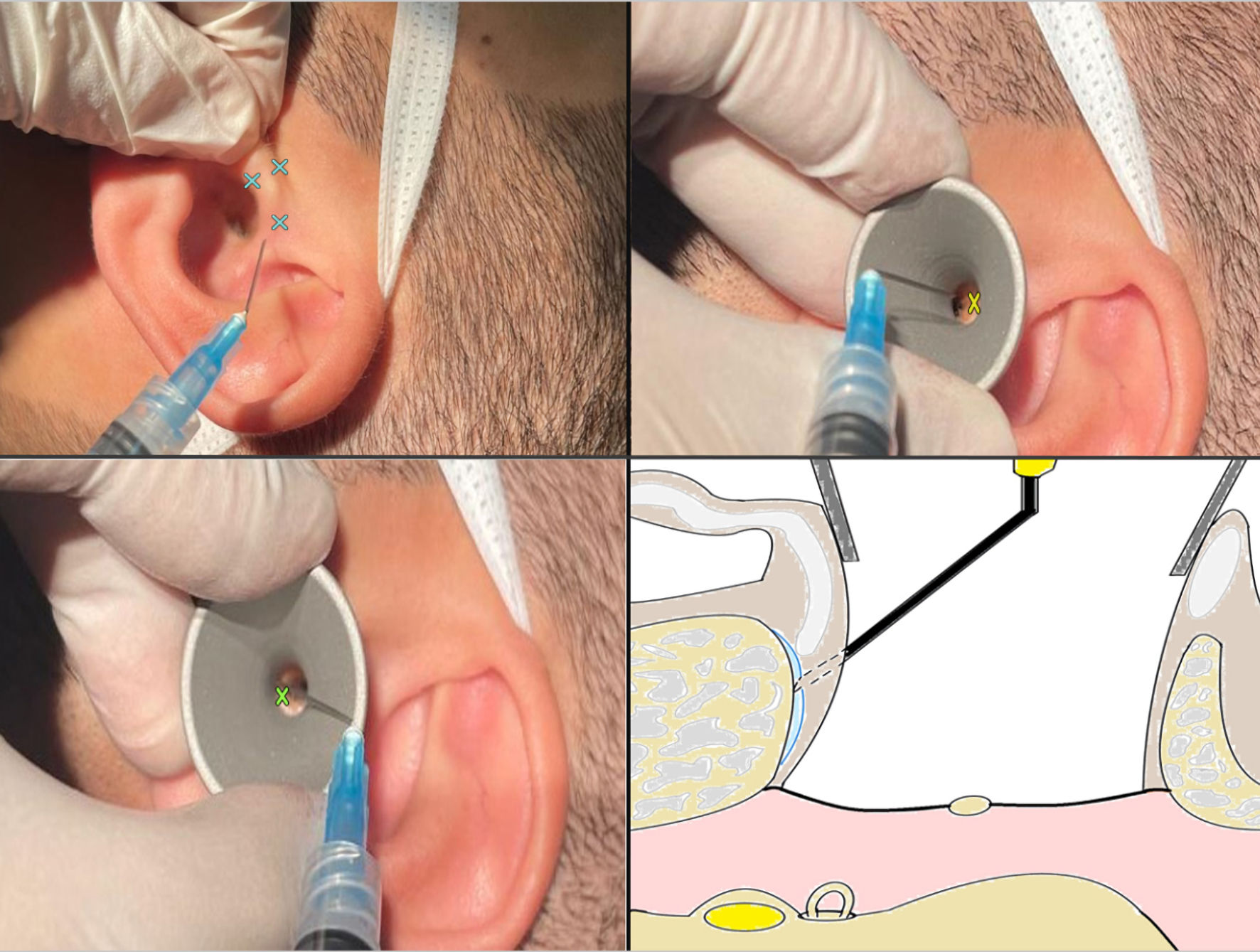
Figure 3.
Figure 3. The patient is in a flat position with no elevation under the head, with the head tilted to the opposite side (yellow arrow) and the chin into their chest (green arrow) to improve the visibility of the middle ear.
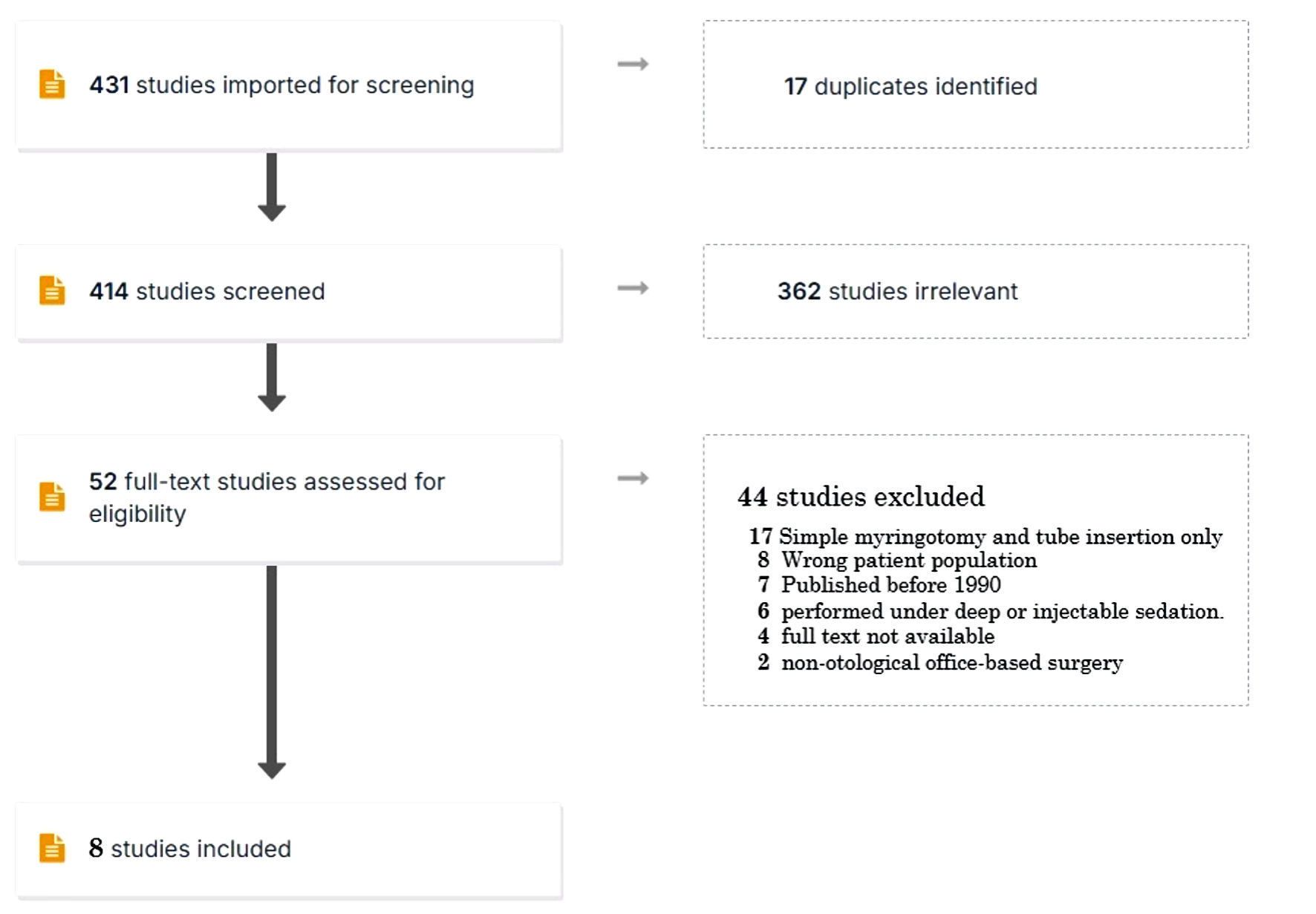
Figure 4.
Figure 4. Tragal cartilage and the external auditory canal infiltration sites (cross marks). The metallic segment of the needle can be bent laterally to provide correct bone contact. Top left: tragal cartilage infiltration sites. Top right: superior part of the external auditory canal infiltration site. Bottom left: inferior part of the external auditory canal infiltration site. Bottom right: correct infiltration depth, the metallic segment of the needle should be bent laterally to ensure proper bone contact.