Figures
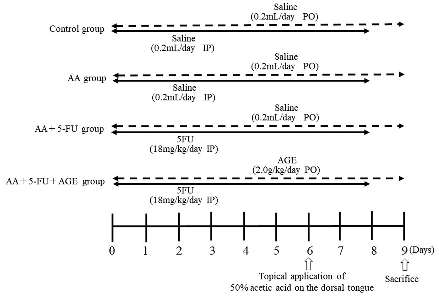
↓ Figure 1. Experimental protocol and schedule.
Three groups (AA, AA + 5-FU, and AA + 5-FU + AGE) are SCCVII tumor-bearing mice (n = 4), and the other
group is control without any tumor (n = 4). Oral mucositis was induced in AA + 5-FU and AA + 5-FU + AGE
groups by intraperitoneal injection of 5-FU (18 mg/kg/day) for 9 days (from day 0 to day 8); while the
control and AA groups received saline (0.2 mL/day) intraperitoneally for the same period. Mice of all
groups except for the control received 50% AA on the dorsal tongue on day 6. As a treatment for oral
mucositis, AA + 5-FU + AGE group received AGE (2.0 g/kg/day) orally for 10 days (day 0 to day 9), while
the control, AA, and AA + 5-FU groups received saline orally (0.2 mL/day) for the same period. All mice
were sacrificed on day 9. AA: acetic acid; AGE: aged garlic extract; 5-FU: 5-fluorouracil.
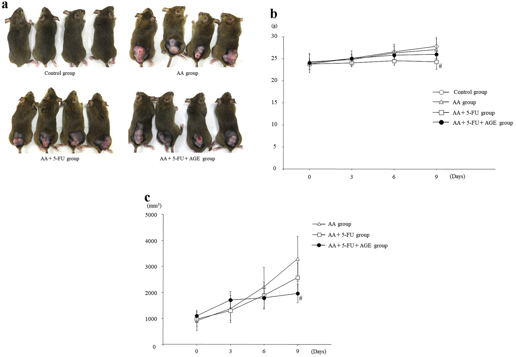
↓ Figure 2. Effect of AGE on body weight and
tumor volume in mice. (a) Photos of the control and tumor-bearing mice taken just before sacrifice. (b)
Graph showing average body weight (g) of mice in each group. 5-FU treatment caused weight loss in AA +
5-FU group and AA + 5-FU + AGE group compared to the control. However, the average body weight was
higher in AA + 5-FU + AGE group than in AA + 5-FU group. Briefly, AGE might have improved mouse health.
Mean ± SEM; #P < 0.05 (two-way analysis of variance and Tukey-Kramer post hoc
test). (c) Graph showing the effect of 5-FU and/or AGE on tumor growth in different groups. Tumor volume
(mm3) was significantly decreased in AA + 5-FU + AGE group compared to the AA + 5-FU group
and AA group. AGE did not negatively influence the antitumor effects of 5-FU, probably enhanced it. Mean
± SEM; #P < 0.05 (two-way analysis of variance and Tukey-Kramer multiple post
hoc test). AA: acetic acid; AGE: aged garlic extract; 5-FU: 5-fluorouracil; SEM: standard error
of the mean.
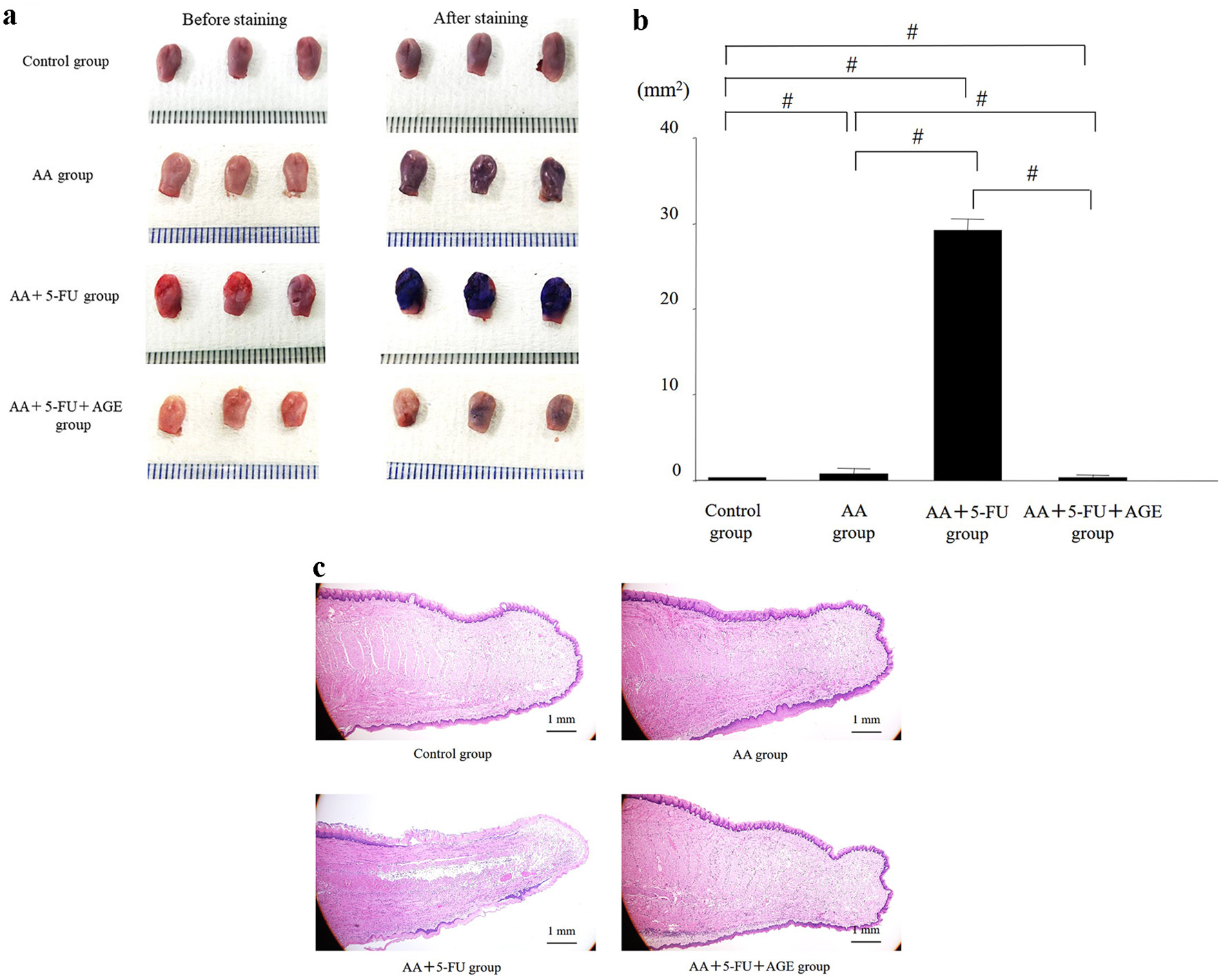
↓ Figure 3. Effect of AGE on the
wounded/mucositis area or ulcerative lesions on the mouse dorsal tongue. (a) Photos of the mouse tongues
before (left side) and after toluidine blue staining (right side). Toluidine blue stains made wounded
areas or ulcers visible. The largest (toluidine blue- positive) wound areas with strong staining were
observed in AA + 5-FU group. Mildly stained small wounds were visible on the tongues of AA group. The
staining intensity was significantly reduced and the wounds were comparatively smaller in AA + 5-FU +
AGE group than those in AA + 5-FU group. (b) Graph showing the effect of AGE on tongue wound/mucositis
area (mm2) in different groups. The toluidine blue-positive tongue wound area was the largest
in AA + 5-FU group. The wound area in AA + 5-FU + AGE group was significantly smaller compared to all
other groups. AGE treatment could heal the wound/ulcer caused by 5-FU and AA. Mean ± SEM;
#P < 0.05 (one-way analysis of variance and Tukey-Kramer post hoc test). (c)
H&E staining of the tongue sections. Severe damage in the epithelial region was observed in AA +
5-FU group, while epithelial damage was almost absent in AA + 5-FU + AGE group. AGE could protect and
heal tongues from 5-FU and AA-induced damage. AA: acetic acid; AGE: aged garlic extract; 5-FU:
5-fluorouracil; H&E: hematoxylin and eosin; SEM: standard error of the mean.
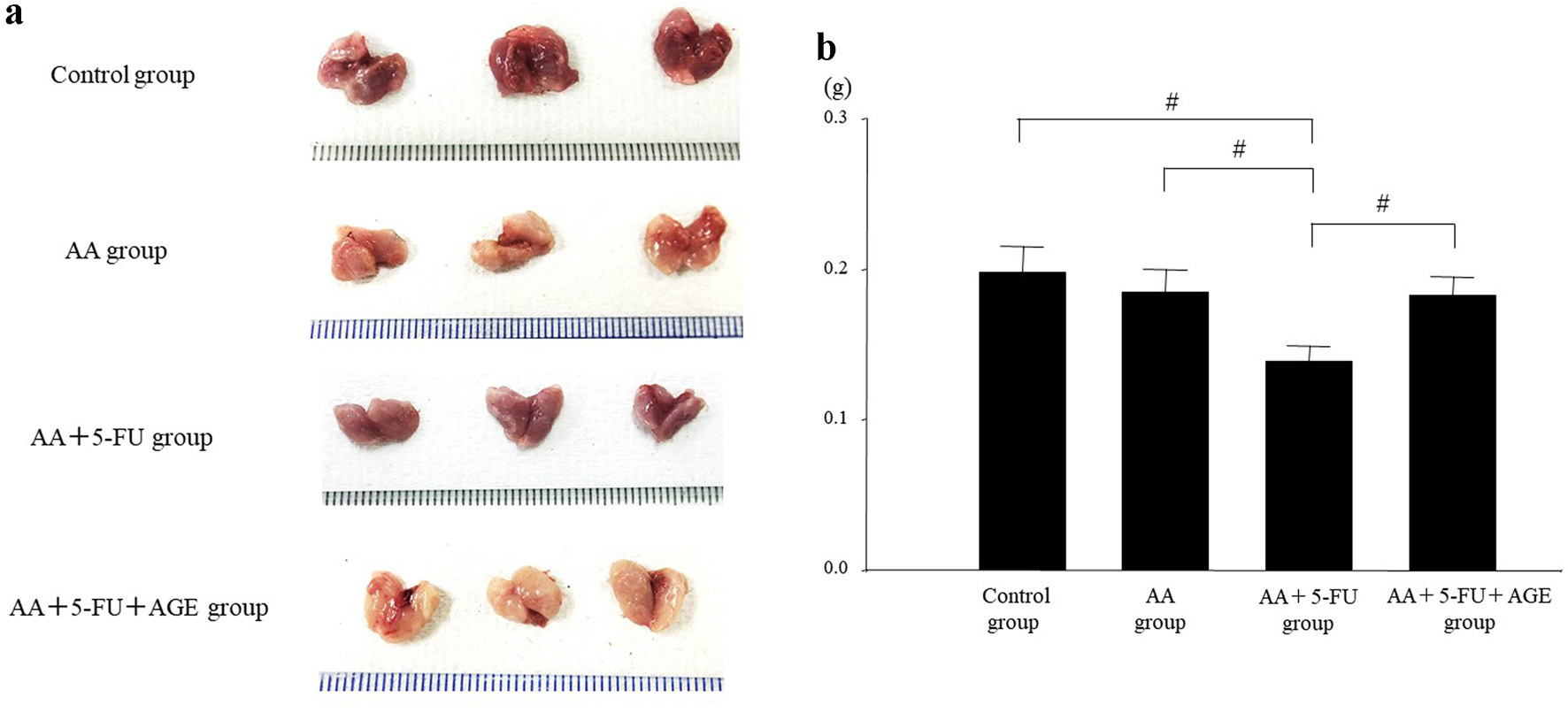
↓ Figure 4. Effect of AGE on the size and weight
of salivary glands in mice. (a) Photos of the extracted salivary glands at the time of sacrifice.
Salivary gland size was smaller in the all treatment groups (AA, AA + 5-FU, and AA + 5-FU + AGE)
compared to the control. The gland size was the smallest in AA + AGE group, while it was increased
markedly in AA + 5-FU + AGE group compared to AA + 5-FU group. (b) Graph showing the effect of AGE on
salivary gland weight (g) in different groups. There was no significant difference in the average weight
of the salivary glands between the control and AA group. Salivary gland weight was significantly
decreased in AA + 5-FU group, which was recovered in AA + 5-FU + AGE group after AGE treatment. Mean
± SEM; #P < 0.05 (one-way analysis of variance and Tukey-Kramer post hoc
test). AA: acetic acid; AGE: aged garlic extract; 5-FU: 5-fluorouracil; SEM: standard error of the
mean.
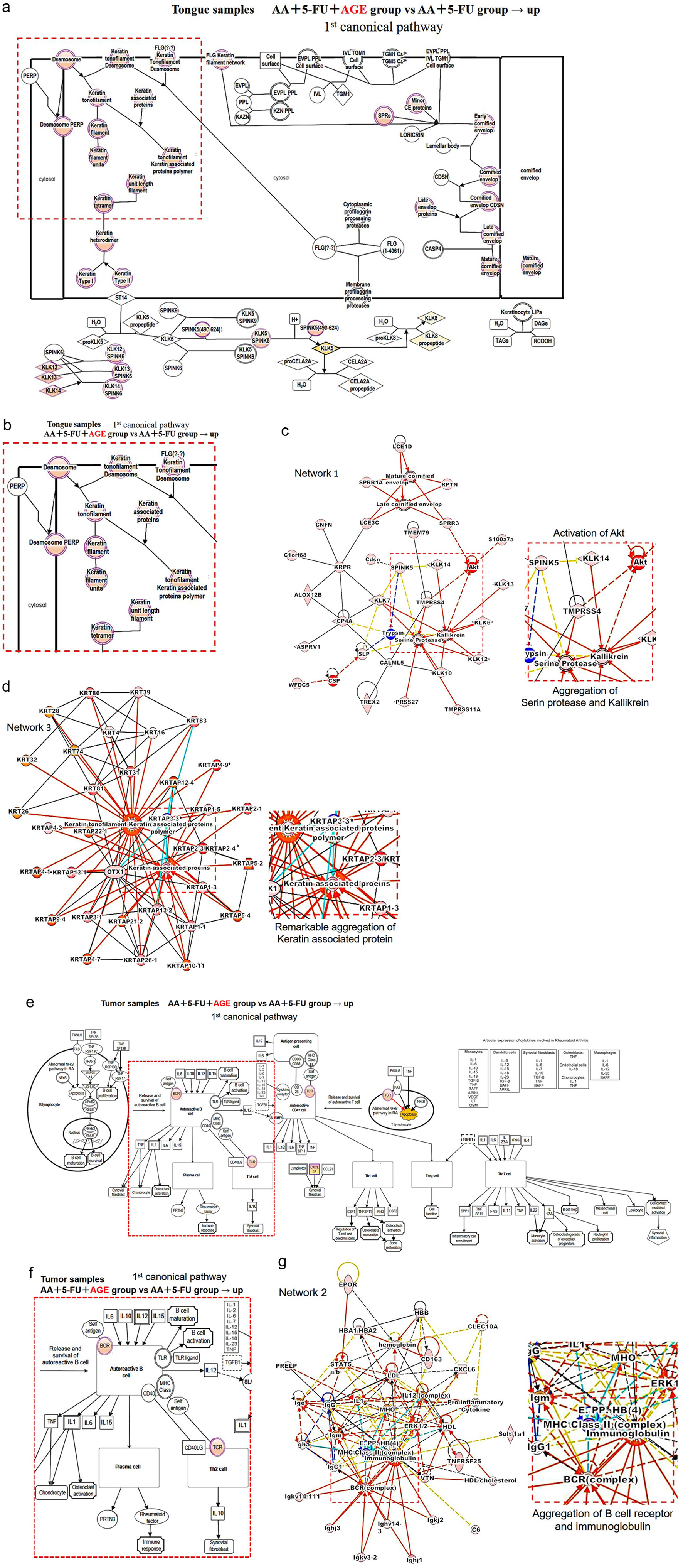
↓ Figure 5. Mechanism of action of wound healing
and antitumor effect of AGE. (a) The first canonical pathway for tongue samples. The genes marked in
pink showed up-regulated expression between these two groups: AA + 5-FU + AGE vs. AA + 5-FU. An enlarged
view of the marked area (by a red square) is shown in (b). (b) These “pink” genes were
up-regulated in AA + 5-FU + AGE group, which are related to the keratinization process. (c) Network 1.
The part inside this red square is enlarged on the right side. It shows the activation of Akt and the
subsequent aggregation of serine protease and kallikrein in AA + 5-FU + AGE group. (d) Network 3. The
enlarged view of the red square area is shown on the right side. There was a noticeable aggregation of
keratin-associated proteins in AA + 5-FU + AGE group. (e) The first canonical pathway for tumor samples.
Up-regulated gene expression between the two groups (AA + 5-FU and AA + 5-FU + AGE) is shown by pink
color. An enlarged view of the marked area (by a red square) is shown in (f). (f) BCR and TCR were
up-regulated in AA + 5-FU + AGE group. (g) Network 2. The red square area is enlarged on the right side.
Aggregation of BCR (BCR complex) and immunoglobulin was observed in AA + 5-FU + AGE group. 5-FU:
5-fluorouracil; AA: acetic acid; AGE: aged garlic extract; BCR: B-cell receptor; TCR: T-cell
receptor.