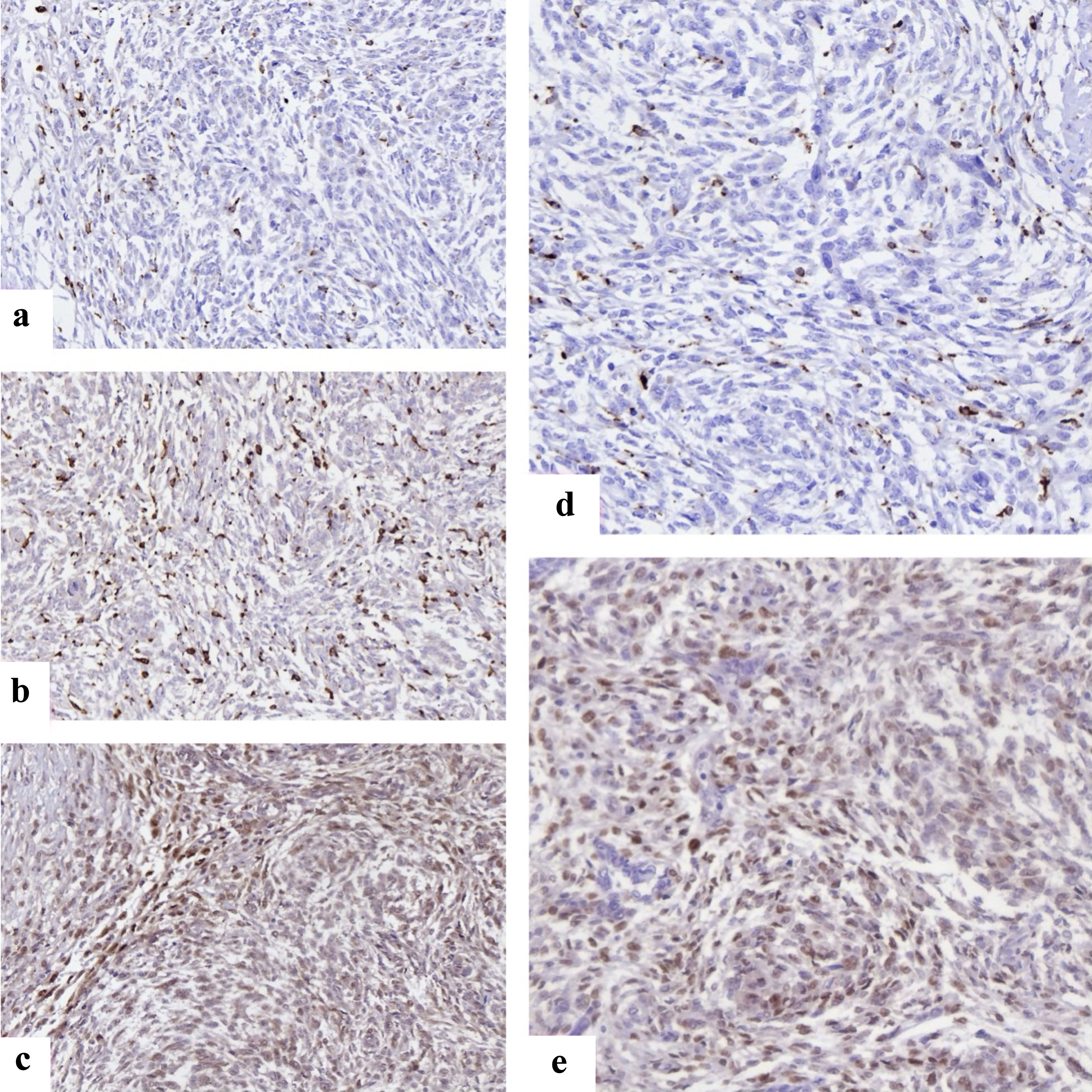
Journal of Clinical Medicine Research, ISSN 1918-3003 print, 1918-3011 online, Open Access
Article copyright, the authors; Journal compilation copyright, J Clin Med Res and Elmer Press Inc
Journal website https://jocmr.elmerjournals.com

Original Article

Volume 000, Number 000, January 2026, pages 000-000


Clinicopathologic and Immunohistochemical Correlates of Disease-Free Survival in Endometrial Stromal Sarcomas: A Multicenter Retrospective Study From 2017 to 2025

Figures

Figure 1.
Figure 1. Representative immunohistochemical staining of immune and cell cycle markers in endometrial stromal sarcomas. (a) FOXP3+ regulatory T cells with distinct nuclear staining scattered within tumor stroma, reflecting immune evasion. (b) CD68 positivity showing widespread macrophage infiltration across tumor areas. (c) CD163 highlights M2-polarized macrophages, enriched at invasive margins, supporting an immunosuppressive microenvironment. (d) Cyclin D1 nuclear overexpression in HG-ESS, consistent with deregulated cell cycle progression. (e) CDK4 nuclear and cytoplasmic expression in HG-ESS tumor cells. CDK4: cyclin-dependent kinase 4; FOXP3: forkhead box P3; HG-ESS: high-grade endometrial stromal sarcoma.
Figure 2.
Figure 2. Kaplan-Meier disease-free survival (DFS) curves stratified by histological group. ESN: endometrial stromal nodule; LG-ESS: low-grade endometrial stromal sarcoma; HG-ESS: high-grade endometrial stromal sarcoma.
Figure 3.
Figure 3. Multivariate analysis of predictors of recurrence in endometrial stromal tumors. ER: estrogen receptor; PR: progesterone receptor; LVSI: lymphovascular space invasion; HG-ESS: high-grade endometrial stromal sarcoma; CI: confidence interval.