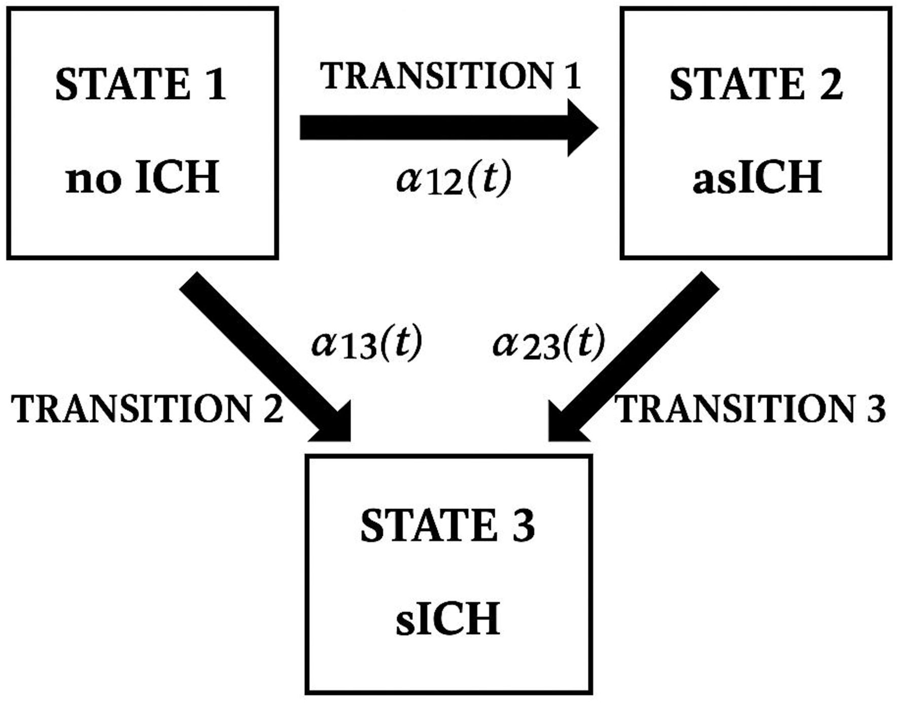
Journal of Clinical Medicine Research, ISSN 1918-3003 print, 1918-3011 online, Open Access
Article copyright, the authors; Journal compilation copyright, J Clin Med Res and Elmer Press Inc
Journal website https://jocmr.elmerjournals.com

Original Article

Volume 17, Number 10, October 2025, pages 556-564


Optimal Timing of Post-Alteplase Brain Computed Tomography: Routine Twenty-Four-Hour Versus Extended Forty-Eight-Hour for Detecting Asymptomatic Intracranial Hemorrhage

Figures

Figure 1.
Figure 1. Conceptual illustration of post-alteplase outcomes and transitions adapted from a multi-state (illness-death) model. ICH: intracranial hemorrhage; asICH: asymptomatic intracranial hemorrhage; sICH: symptomatic intracranial hemorrhage.
Figure 2.
Figure 2. Study flow diagram. Numbers are provided only for groups identified after the first follow-up brain CT scan; subsequent counts are omitted because outcomes evolved dynamically over time and were analyzed using a multi-state (illness-death) model. IV: intravenous; ED: emergency department; MT: mechanical thrombectomy; TOAST: Trial of Org 10172 in Acute Stroke Treatment; CT: computed tomography; ICH: intracranial hemorrhage; asICH: asymptomatic intracranial hemorrhage; sICH: symptomatic intracranial hemorrhage.
Figure 3.
Figure 3. Cumulative incidence of intracranial hemorrhage (ICH) over time following intravenous alteplase administration.

Tables

Table 1. Baseline Characteristics by Final Transitioned Outcomes at 7-Day Follow-Up
 
Baseline characteristicssICH (n = 49)asICH (n = 51)no ICH (n = 455)P value
Numbers in each group represent the final transitioned outcomes at the end of the 7-day follow-up. These differ from those in the study flow diagram, which reflect initial outcomes after the first follow-up brain CT scan. sICH: symptomatic intracranial hemorrhage; asICH: asymptomatic intracranial hemorrhage; ICH: intracranial hemorrhage; SD: standard deviation; NIHSS: National Institutes of Health Stroke Scale; IQR: interquartile range; CT: computed tomography.
Male, n (%)22 (44.9)30 (56.6)252 (55.6)0.225
Age (years), mean ± SD69.7 ± 9.964.4 ± 10.164.5 ± 12.70.416
Body weight (kg), mean ± SD57.5 ± 12.160.9 ± 13.958.3 ± 12.80.809
Initial NIHSS, median (IQR)13 (10, 18)12 (8, 17)10 (7, 13)< 0.001
Number of brain CT scans during admission, median (min, max)2 (1, 6)1 (1, 3)1 (1, 4)< 0.001
Dead during admission, n (%)
  Neurological cause15 (30.6)0 (0)8 (1.8)0.006
  Non-neurological cause0 (0)1 (1.9)7 (1.5)


Table 2. Frequency of Brain CT Scans for Asymptomatic Intracranial Hemorrhage Within 7 Days
 
Follow-up brain CT scans and timing of first detectionAsymptomatic ICH (n = 51)
Data represent patients whose final transitioned outcome was classified as asymptomatic intracranial hemorrhage (asICH) at the end of the 7-day follow-up. CT: computed tomography; ICH: intracranial hemorrhage; SD: standard deviation.
Number of brain CT scans performed
  1-time CT patients, n (%)37 (69.8)
  2-time CT patients, n (%)11 (20.8)
  3-time CT patients, n (%)5 (9.4)
Time to first detection (h), mean ± SD25.7 ± 8.7


Table 3. Comparison of First Asymptomatic Intracranial Hemorrhage (asICH) Detection Between 24-Hour Routine and 48-Hour Extended Brain Computed Tomography Scenarios
 
First transition to asymptomatic intracranial hemorrhage (asICH) (n = 63a)
CT timing scenario 24-h routineCT timing scenario 48-h extendedP value
aN = 63 represents the first detection of asymptomatic intracranial hemorrhage (asICH), which differs from the final transitioned outcome (n = 51), as 12 patients progressed to symptomatic intracranial hemorrhage (sICH) within 7 days. CT: computed tomography.
First asICH detection, n (%) - sensitivity19 (30.2)61 (96.8)< 0.001