Journal of Clinical Medicine Research, ISSN 1918-3003 print, 1918-3011 online, Open Access
Article copyright, the authors; Journal compilation copyright, J Clin Med Res and Elmer Press Inc
Journal website https://jocmr.elmerjournals.com

Original Article

Volume 18, Number 5, May 2026, pages 336-342


Perioperative and Long-Term Outcomes of T-Shaped Esophagojejunostomy After Laparoscopic Total Gastrectomy for Gastric Cancer

Figures

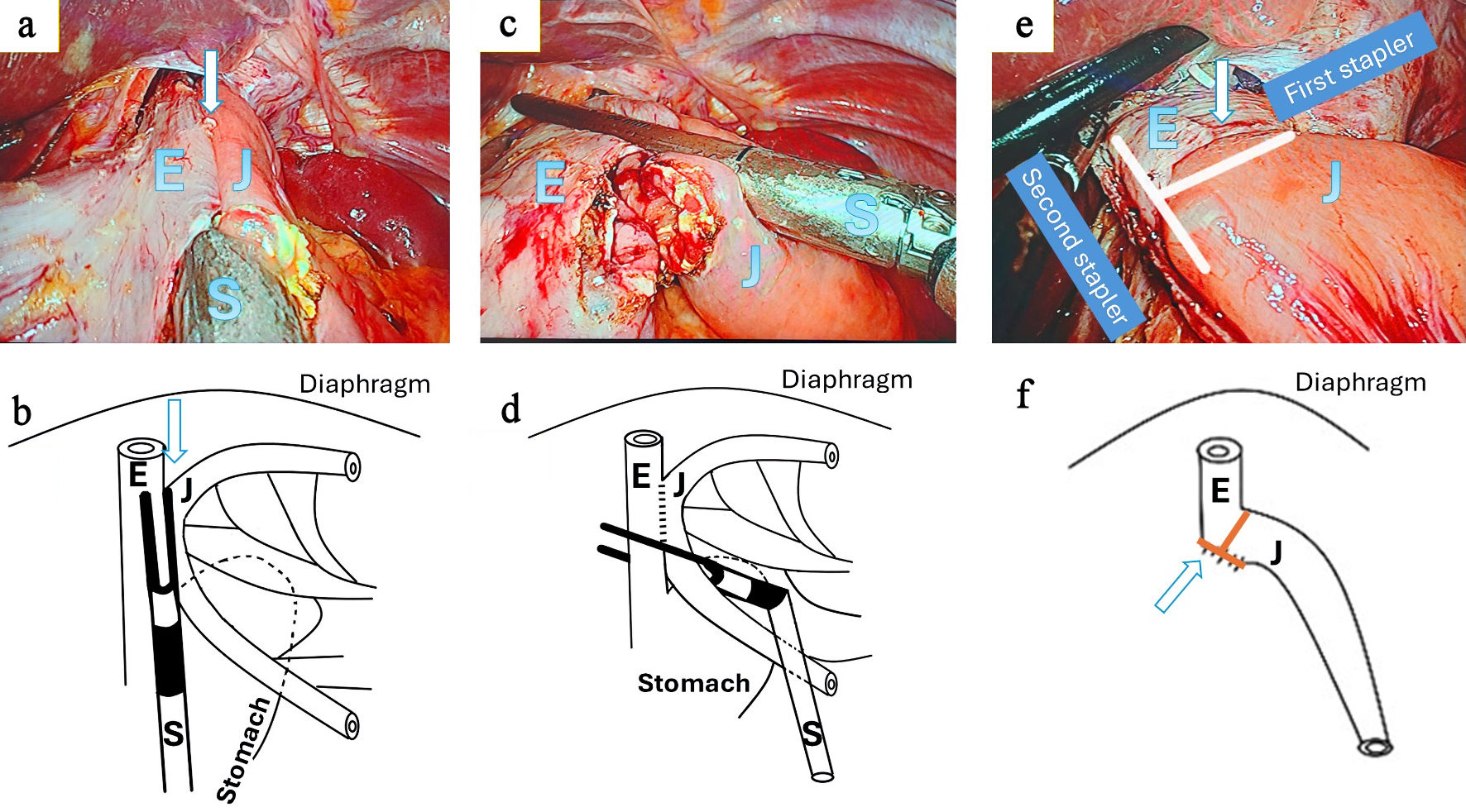
↓  Figure 1. Intracorporeal T-shaped functional end-to-end esophagojejunostomy. (a, b) Creation of the initial side-to-side esophagojejunostomy. Small enterotomies are made on the lateral wall of the esophagus (E) and the antimesenteric border of the jejunum (J), followed by insertion and firing of a linear stapler (S) to create a functional side-to-side anastomosis (arrow). (c, d) Perpendicular stapling step. A second linear stapler is applied perpendicular to the initial staple line, incorporating both the esophageal and jejunal walls at the level of the common entry hole. Upon firing, this step closes the common entry hole and trims the overlapping tissue edges rather than resecting the anastomosis. (e, f) Final configuration of the T-shaped functional end-to-end esophagojejunostomy. The intersection of the two staple lines creates a characteristic T-shaped geometry (arrow), resulting in a straight luminal alignment without a residual blind pouch or jejunal stump. The apparent stump-like structure corresponds to the transverse staple line rather than a true jejunal stump. E: esophagus; J: jejunum; S: stapler.
Figure 1.
↓  Figure 2. Postoperative contrast study. Contrast study shows a smooth and continuous luminal passage from the esophagus (E) to the jejunum (J), without evidence of a blind pouch or diverticular formation. The arrow indicates the anastomotic site.
Figure 2.

Tables

↓  Table 1. Baseline Characteristics of the Study Population (N = 167)
 
Age (years)60.97 ± 12.05 (range: 26–88)
Values are presented as mean ± standard deviation or n (%), unless otherwise indicated. Pathological stage was defined according to the Japanese Gastric Cancer Association (JGCA) Classification, Fifth Edition. ASA: American Society of Anesthesiologists physical status classification.
BMI (kg/m2)18.07 ± 6.40
Sex
  Male118 (70.7%)
  Female49 (29.3%)
ASA physical status
  I71 (42.5%)
  II73 (43.7%)
  III23 (13.8%)
Tumor location
  Upper third20 (12.0%)
  Middle third141 (84.4%)
  Diffuse infiltrative6 (3.6%)
Histological type
  Tubular or papillary adenocarcinoma99 (59.3%)
  Mucinous adenocarcinoma36 (21.5%)
  Signet-ring cell carcinoma32 (19.2%)
Histological differentiation
  Well differentiated38 (22.8%)
  Moderately differentiated51 (30.5%)
  Poorly/undifferentiated78 (46.7%)
Pathological stage (JGCA)
  IA18 (10.8%)
  IB20 (12.0%)
  IIA43 (25.7%)
  IIB33 (19.8%)
  IIIA14 (8.4%)
  IIIB31 (18.6%)
  IIIC8 (4.8%)


↓  Table 2. Operative Outcomes (N = 167)
 
Technical feasibility
Values are presented as mean ± standard deviation or n (%), unless otherwise indicated. Estimated blood loss was calculated intraoperatively by the anesthesiology team. Technical errors during anastomosis were defined as intraoperative stapler misfiring or need for additional suturing without clinical sequelae. D2 lymphadenectomy was performed according to the Japanese Gastric Cancer Association guidelines.
  Estimated blood loss (mL)34.04 ± 14.30
  Conversion to open surgery0 (0%)
  Nasojejunal tube placement62 (37.2%)
  Operative time (min)207.01 ± 36.0 (range: 120–300)
Oncologic quality
  Retrieved lymph nodes (n)22.95 ± 6.45 (range: 15–45)
  Metastatic lymph nodes (n)2.40 ± 2.75 (range: 0–12)
  Proximal resection margin (cm)5.2 ± 0.9 (range: 2.5–7.0)
  Negative proximal and distal margins167 (100%)
Intraoperative complications
  Splenic vessel injury3 (1.8%)
  Splenic parenchymal injury4 (2.4%)
  Hepatic parenchymal injury3 (1.8%)
  Middle colic artery injury1 (0.6%)
  Small bowel serosal injury1 (0.6%)
  Technical errors during anastomosis3 (1.8%)


↓  Table 3. Postoperative and Long-Term Outcomes (N = 167)
 
Postoperative complications
Values are presented as mean ± standard deviation or n (%), unless otherwise indicated. Postoperative complications were defined as events occurring within 30 days after surgery. Pain intensity was assessed using the visual analog scale (VAS). Overall survival was estimated using the Kaplan–Meier method.
  Pneumonia1 (0.6%)
  Residual intra-abdominal abscess1 (0.6%)
  Surgical site infection2 (1.2%)
  Anastomotic leakage0 (0%)
Postoperative pain (VAS)
  Mild77 (44.5%)
  Moderate72 (41.6%)
  Severe18 (10.4%)
  Very severe0 (0.0%)
Postoperative recovery
  Duration of nasojejunal tube placement (h)92.43 ± 55.6 (range: 24–360)
  Time to drain removal (days)3.7 ± 1.6 (range: 2–7)
  Time to first flatus (h)49.58 ± 16.7 (range: 24–96)
  Time to oral intake (days)3.01 ± 1.09 (range: 2–6)
  Postoperative hospital stay (days)7.45 ± 1.89 (range: 5–15)
Adjuvant chemotherapy
  Yes129 (77.2%)
  No38 (22.8%)
Long-term outcomes
  Mean follow-up duration (months)44.10 ± 20.01 (range: 2–84)
  Postoperative reflux symptoms33 (19.8%)
  Anastomotic stricture3 (1.8%)
  Postoperative bowel obstruction2 (1.8%)
  Anastomotic recurrence1 (0.6%)
  Distant metastasis31 (18.6%)
  Time to metastasis (months)22.5 ± 9.1 (range: 8–52)
Mean overall survival (months)54.38 ± 1.98 (range: 31.15–56.85)