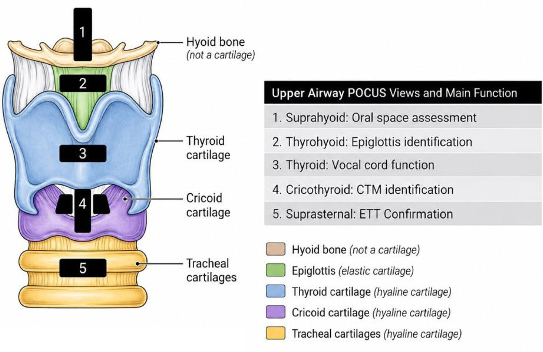
| Kundra et al [5] | Review article | Educational review outlining the use of ultrasound (US) in various aspects of airway management including identification of the difficult airway as well as confirmation of endotracheal tube (ETT) placement, prediction of post-extubation stridor, evaluation of soft tissue masses in the neck, and assessment of subglottic diameter for ETT size. |
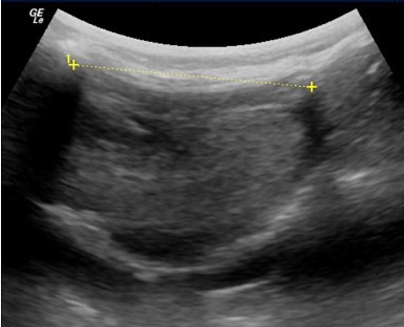
| Carsetti et al [15] | Meta-analysis of 15 studies using airway ultrasound to predict difficult intubation. | Airway US index tests are significantly different between patients with easy versus difficult direct laryngoscopy. Distance from the skin to epiglottis (DSE) is the most studied index test in the literature. |
| Singh et al [35] | Systematic review and meta-analysis of 21 studies in adults (7 airway and 14 non-airway) with 3,339 patients. | Obstructive sleep apnea (moderate-severe) correlated to a moderate degree with the distance between lingual arteries, resting tongue thickness, and tongue base thickness. The non-airway parameter of carotid intimal media thickness had a low to moderate correlation with moderate to severe OSA. |
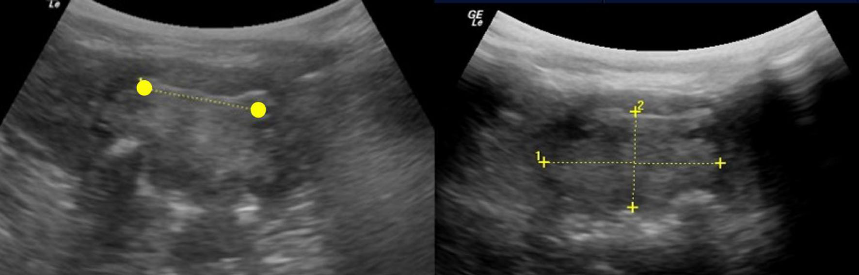
| Husein et al [57] | A prospective, double-blinded pediatric study comparing US, video bronchoscopy, and ETT sizing for measurement of subglottic airway diameter. | US measurements correlated with video-bronchoscopy and ETT sizing. However, US underestimated absolute subglottic dimensions. Despite the authors being less enthusiastic about use of US for absolute airway sizing or ETT selection, they opined that it may serve as a useful non-invasive tool for longitudinal monitoring of changes in subglottic caliber over time. |
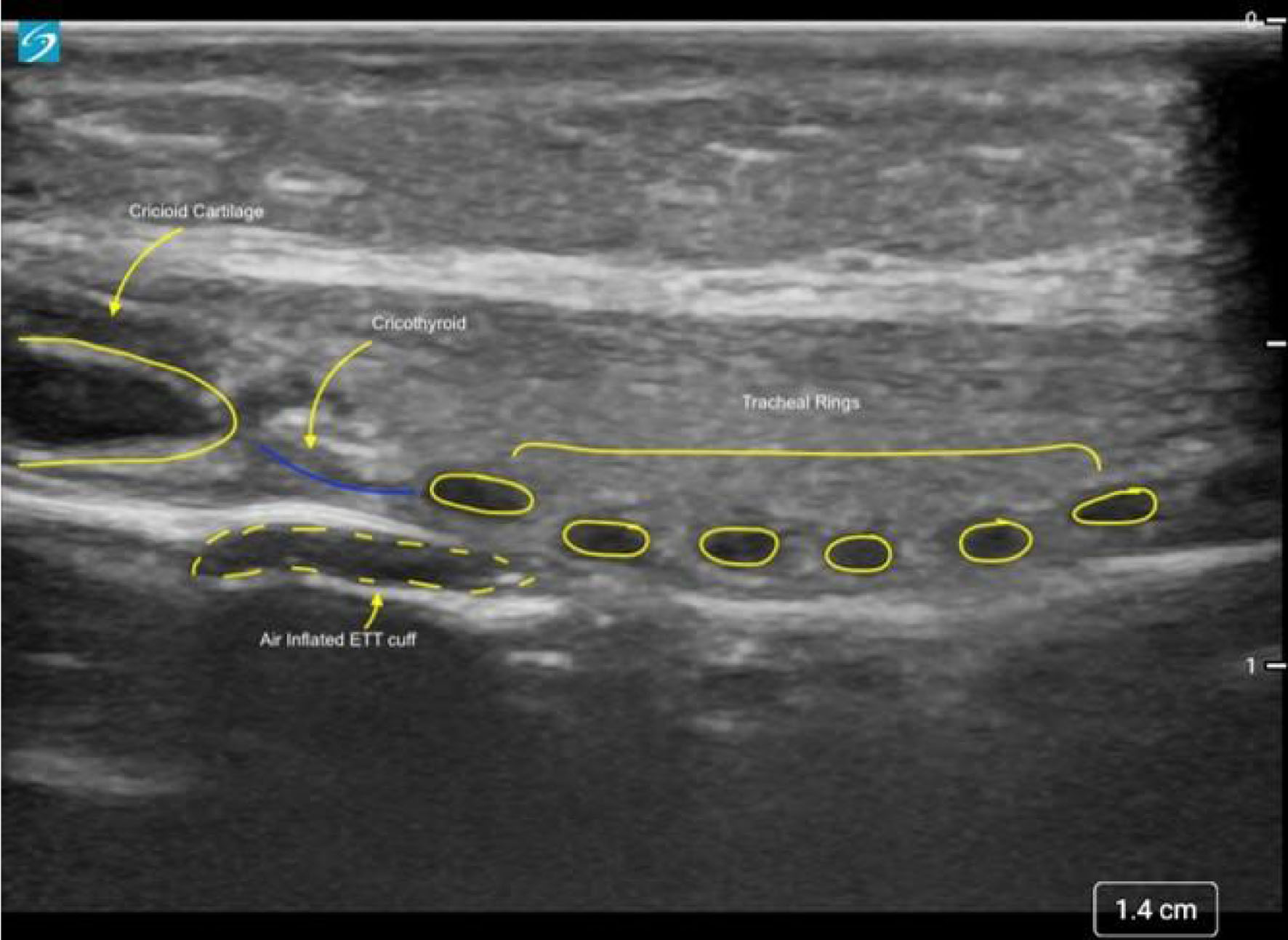
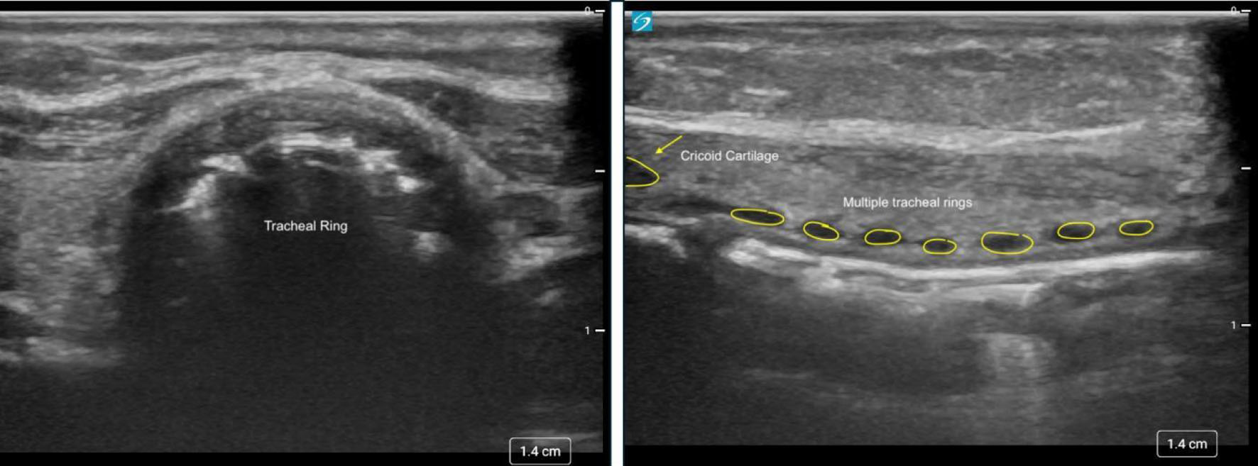
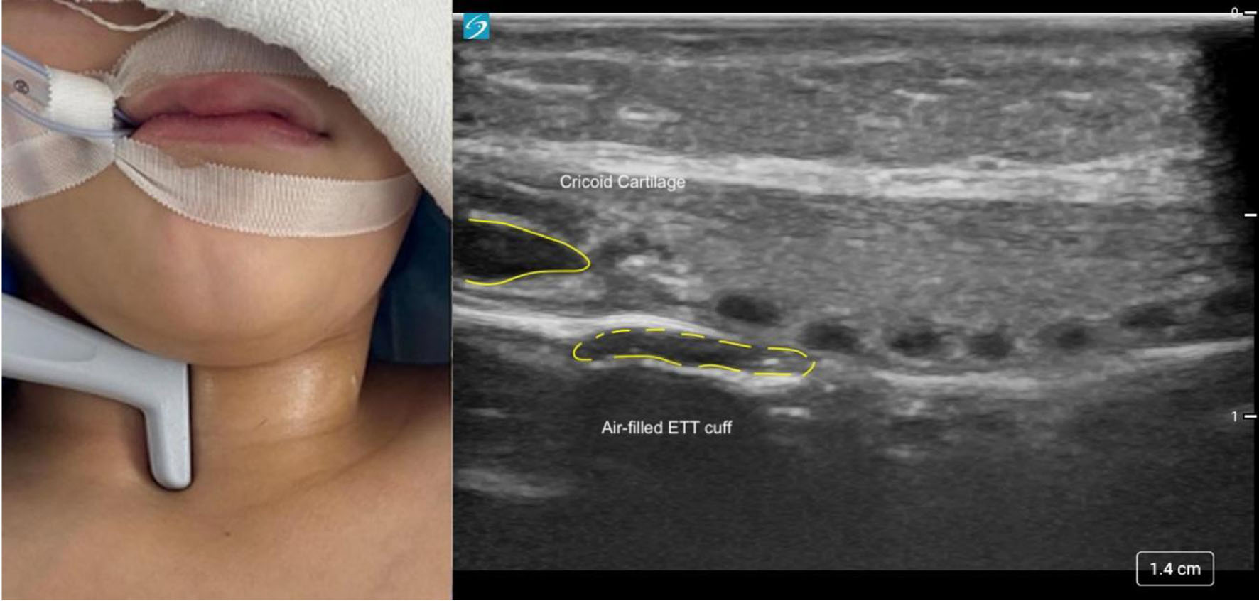
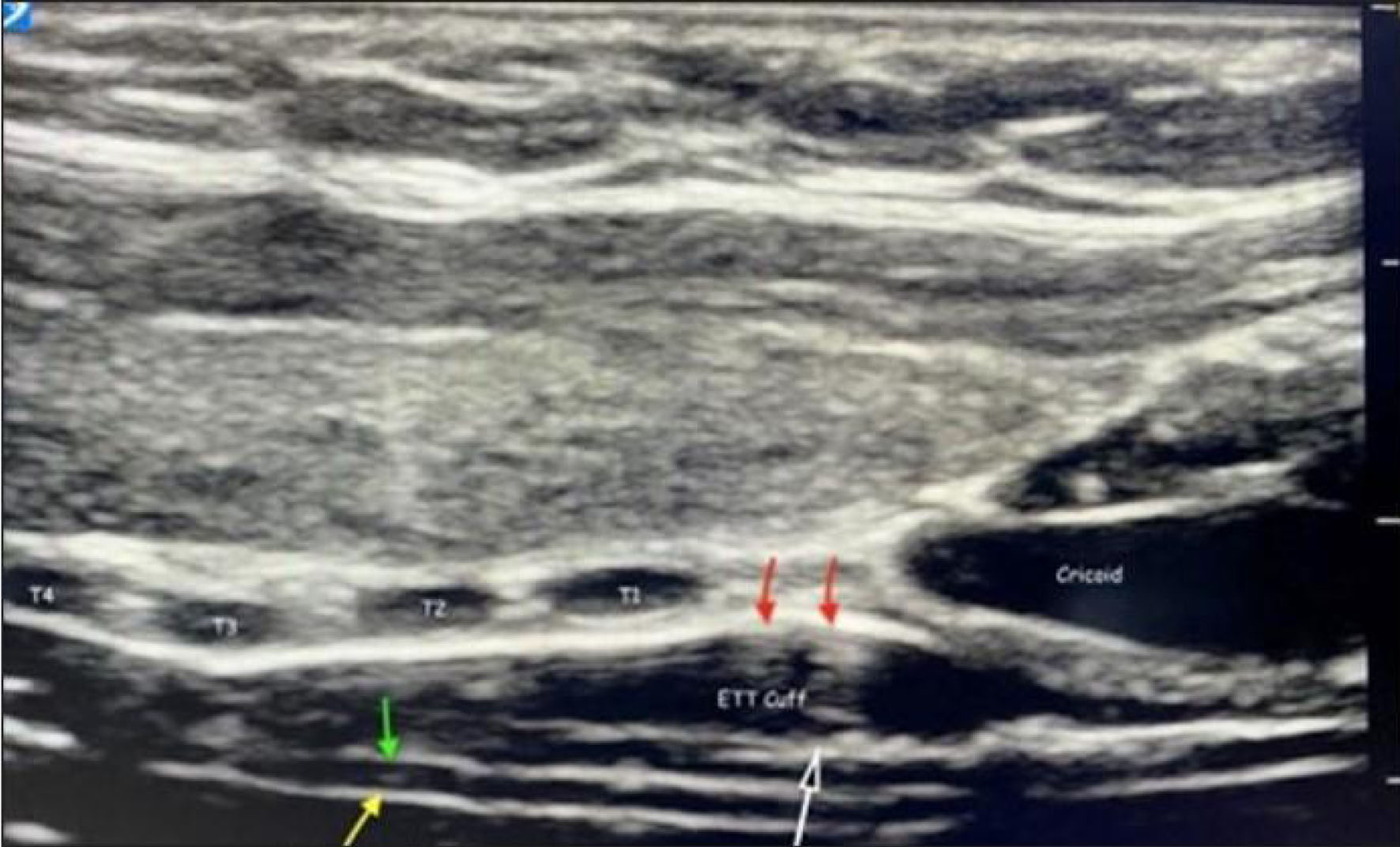
| Wani et al [61] | Prospective, non-randomized study of 80 pediatric patients (1–78 months of age). | US used to locate the ETT cuff relative to the cricoid and tracheal rings. The cephalad end of the ETT cuff was found at the level of the cricoid in 16.3% of patients, at the first, second, and third tracheal rings in 27.5%, 23.8%, and 17.5% of patients, respectively, and at or below the 4th tracheal ring in 15% of patients. The authors suggested that this observed inconsistency highlights known variability in pediatric airway anatomy, underscoring the value of real-time US imaging to document cuff position. |
| Singh et al [68] | Prospective study of 100 pediatric patients (12–60 months of age). | Using US, tracheal diameter was measured as the transverse air column diameter at the cephalad end of the cricoid cartilage. US correlated more strongly with actual ETT size than other formulas (age-based, body length, or finger width). |
| Marciniak et al [80] | Prospective study of 30 pediatric patients (mean age 48 months). | US imaging was used to visualize passage of the ETT into the airway by visualization and identification of the trachea, tracheal rings, and the vocal cords. The authors noted widening of glottis as the ETT passed through the airway and correctly positioned the ETT by using lung sliding. A single case of esophageal intubation was readily recognized by visualization of the ETT in the esophagus in the left paratracheal space. |
| Kerrey et al [82] | Lung US versus chest radiography in 127 intubated pediatric patients. | Chest radiography identified the ETT in the mainstem bronchus in 24 patients, while there were no esophageal intubations. US and chest radiography agreed on ETT placement in 106 patients (94 tracheal and 12 mainstem), for an overall agreement of 0.83. Although diaphragmatic US was not equivalent to chest radiography for ETT placement within the airway, the results were timelier, detected more misplacements than standard clinical confirmation alone, and were highly reproducible. |
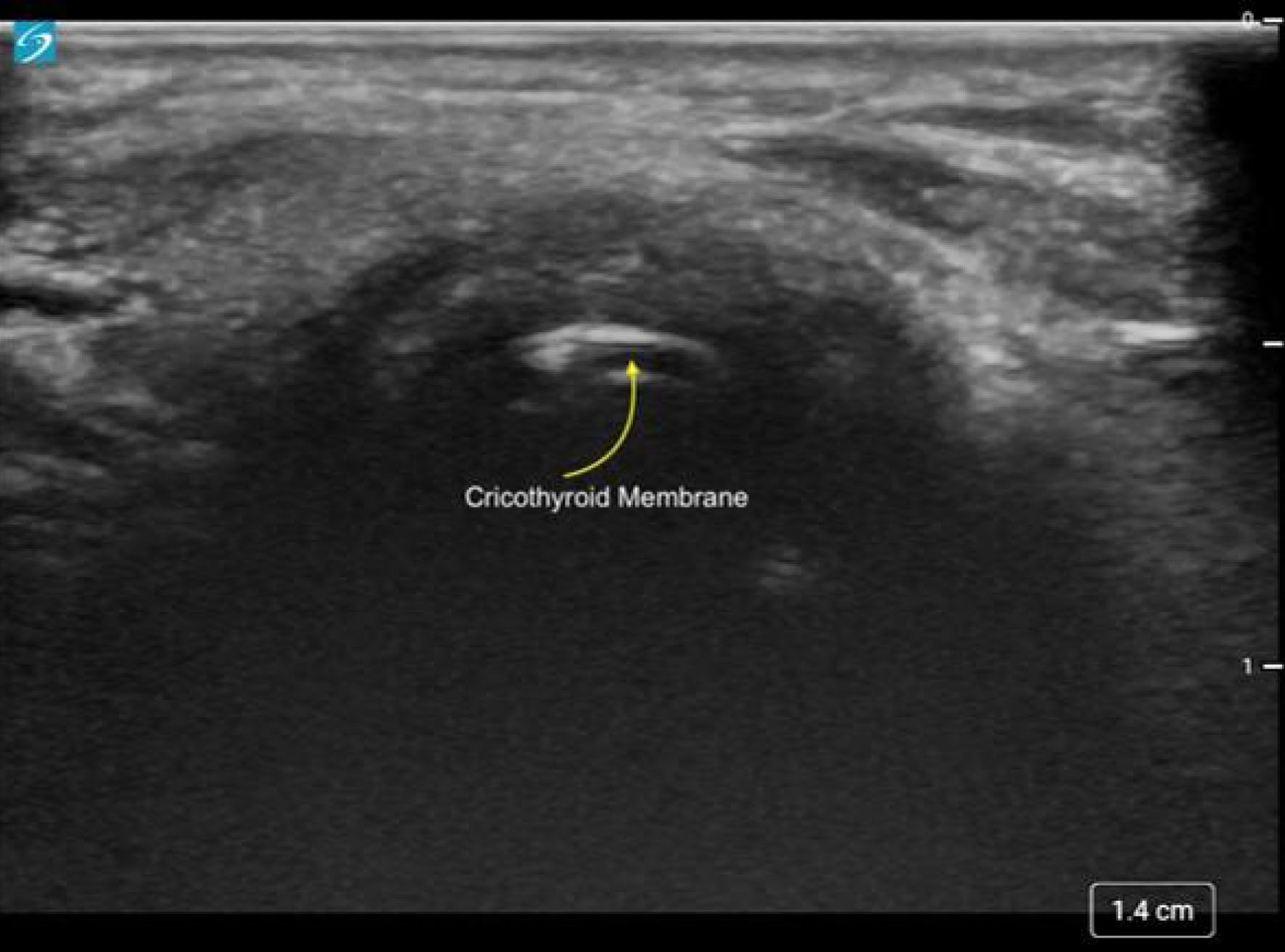
| Walsh et al [84] | Prospective study of 22 pediatric patients. | The cricothyroid membrane (CTM) was accurately identified by US in all cases. Height of the CTM correlated with height measured by MRI. |
| Moharir et al [97] | Prospective, blinded study comparing US with auscultation during one-lung ventilation (OLV) in patients 0–20 years of age. | Lung US demonstrated a diagnostic accuracy of 95%, significantly higher than auscultation (68%, P < 0.001). Although US required a slightly longer assessment time, it provided substantially greater diagnostic reliability, supporting its role as a superior adjunct to auscultation to document effective lung isolation. |