Figures
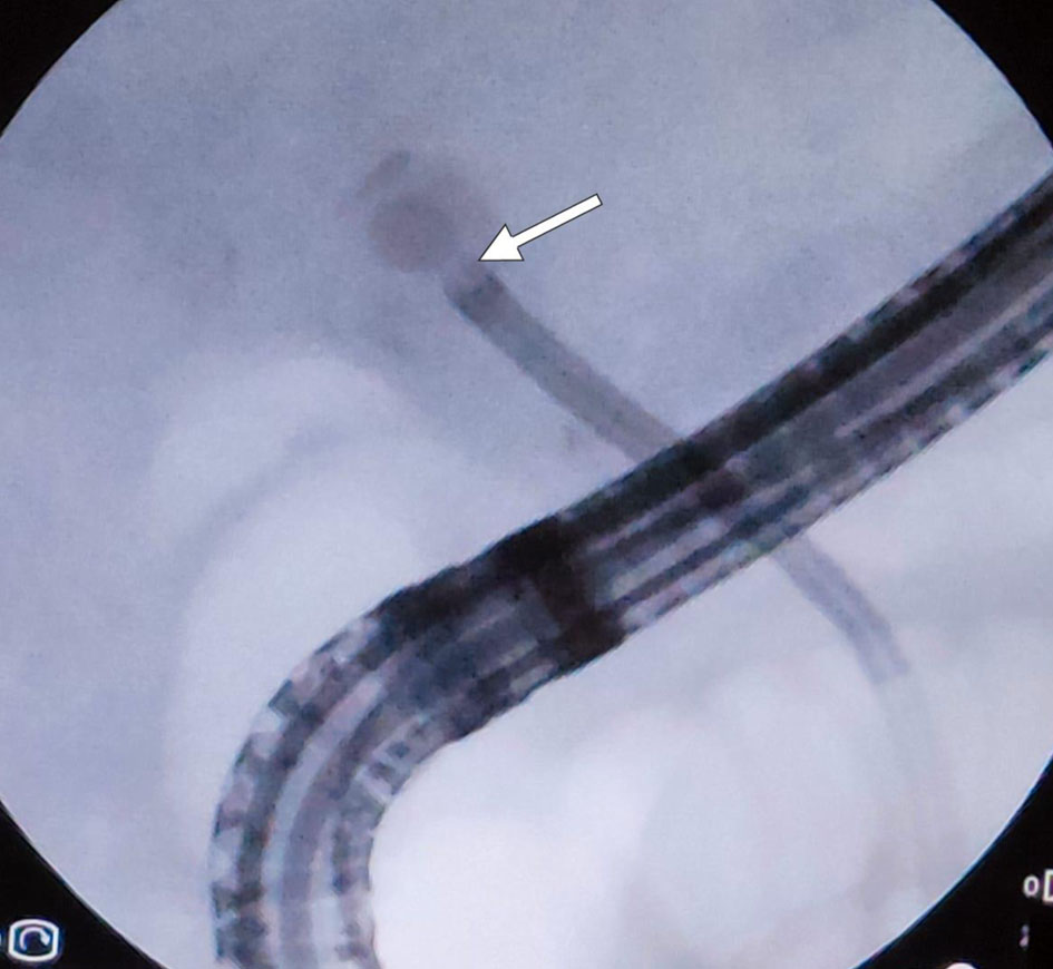
↓ Figure 1. Fluoroscopic image obtained during ERCP demonstrating the single-operator cholangioscope (SpyGlass DS; Boston Scientific, Marlborough, MA, USA) positioned in direct contact with the target CBD stone; the arrow indicates the tip of the cholangioscope abutting the stone surface. CBD: common bile duct; ERCP: endoscopic retrograde cholangiopancreatography.
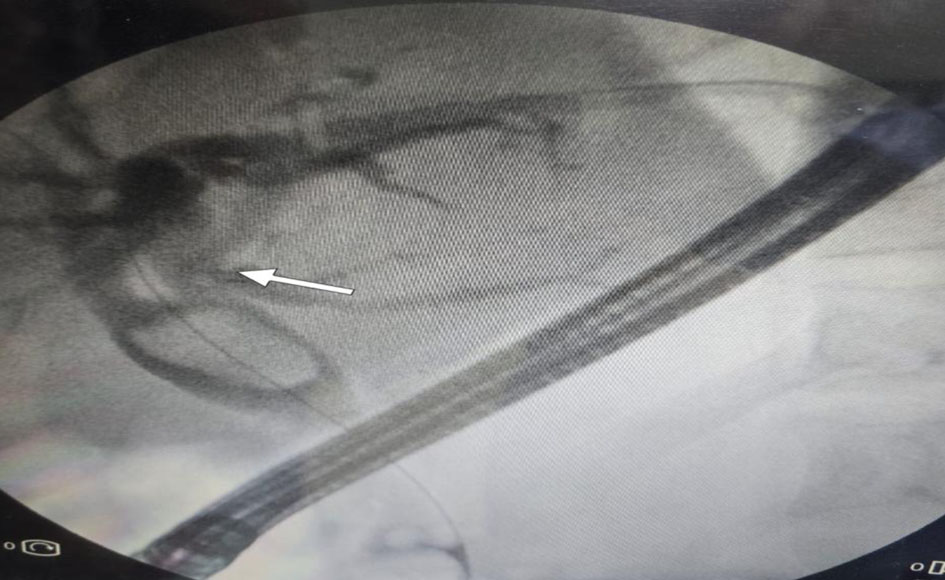
↓ Figure 2. Fluoroscopic cholangiogram showing a large filling defect within the CBD, consistent with a difficult CBD stone; the arrow marks the stone’s proximal margin. CBD: common bile duct.
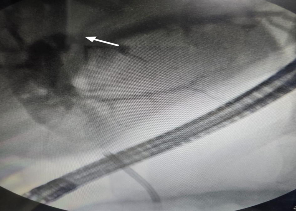
↓ Figure 3. SpyGlass DS single-operator cholangioscope advanced into the CBD to perform EHL. The arrow indicates the cholangioscope tip within the ductal lumen. CBD: common bile duct; EHL: electrohydraulic lithotripsy.
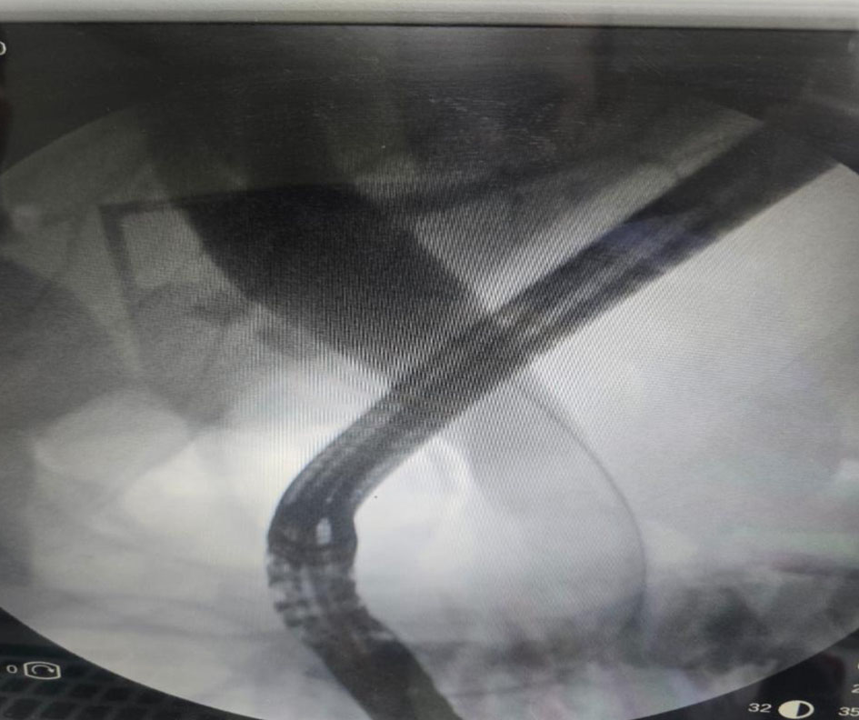
↓ Figure 4. Occlusion cholangiogram, obtained after SOC-guided EHL, confirming complete clearance of the CBD with no residual filling defect. CBD: common bile duct; EHL: electrohydraulic lithotripsy; SOC: single-operator cholangioscopy.
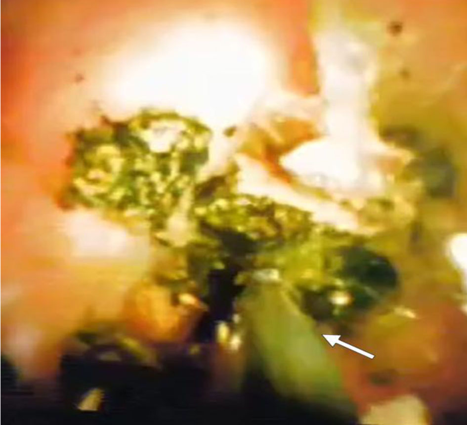
↓ Figure 5. Direct cholangioscopic view during EHL probe lithotripsy, showing the EHL probe (arrow) applied to the stone surface under direct visualization through the single-operator cholangioscope. EHL: electrohydraulic lithotripsy.
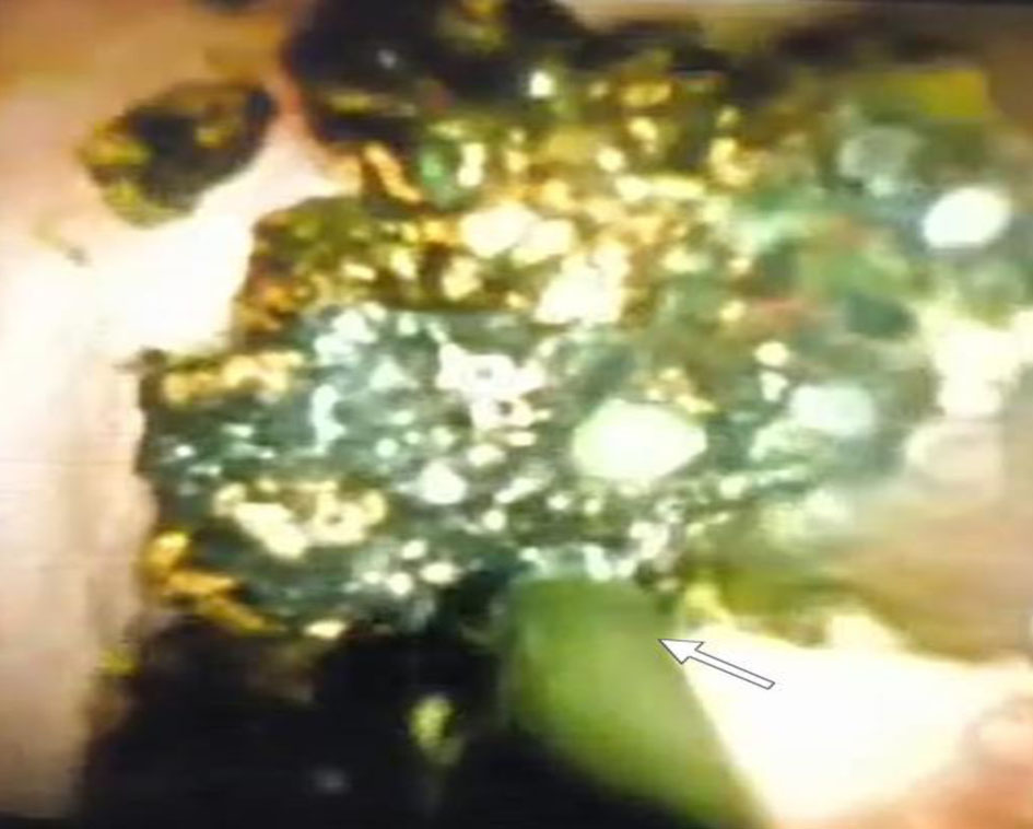
↓ Figure 6. Cholangioscopic image showing fragments of stones in front of EHL probe (arrow), generated by EHL immediately prior to extraction with a balloon sweeper or Dormia basket. EHL: electrohydraulic lithotripsy.
Tables
↓ Table 1. Demographic and Clinical Characteristics of the Study Population (n = 25)
| Characteristic | Value (n = 25) |
|---|
| ERCP: endoscopic retrograde cholangiopancreatography; HCV: hepatitis C virus; RUQ: right upper quadrant; SD: standard deviation; SVR: sustained virological response. |
| Age (years), mean ± SD | 61.8 ± 6.4 |
| Age range (years) | 47–75 |
| Sex, n (%) | |
| Male | 11 (44.0) |
| Female | 14 (56.0) |
| Comorbidities, n (%) | |
| None | 15 (60.0) |
| Hypertension | 7 (28.0) |
| Diabetes mellitus | 6 (24.0) |
| HCV (treated with SVR) | 3 (12.0) |
| Ischemic heart disease | 1 (4.0) |
| Bronchial asthma | 1 (4.0) |
| Clinical presentation, n (%) | |
| RUQ abdominal pain | 16 (64.0) |
| Dyspepsia | 9 (36.0) |
| Epigastric pain | 8 (32.0) |
| Jaundice | 4 (16.0) |
| Fever | 2 (8.0) |
| Dark urine | 4 (16.0) |
| Pale stool | 4 (16.0) |
| Nausea/vomiting | 3 (12.0) |
| Gallbladder status, n (%) | |
| Post-cholecystectomy | 16 (64.0) |
| Gallbladder in situ | 9 (36.0) |
| Previous ERCP, n (%) | |
| No previous ERCP | 1 (4.0) |
| At least one prior ERCP | 24 (96.0) |
↓ Table 2. Pre-Procedural Laboratory Parameters of the Study Population (n = 25)
| Parameter | Mean ± SD | Reference range | P-value |
|---|
| ALP: alkaline phosphatase; ALT: alanine aminotransferase; AST: aspartate aminotransferase; CRP: C-reactive protein; ESR: erythrocyte sedimentation rate; GGT: gamma-glutamyl transferase; INR: international normalized ratio; SD: standard deviation. |
| Hemoglobin (g/dL) | 12.68 ± 1.51 | 12.0–16.0 | < 0.001 |
| Total leukocyte count (× 103/µL) | 7.26 ± 2.27 | 4.0–11.0 | 0.602 |
| Platelet count (× 103/µL) | 269.20 ± 97.47 | 150–400 | 0.769 |
| ESR (mm/h) | 36.92 ± 8.72 | 0–20 | < 0.001 |
| CRP (mg/L) | 10.00 ± 14.04 | 0–6 | 0.020 |
| INR | 1.04 ± 0.09 | 0.8–1.2 | 0.036 |
| AST (U/L) | 29.92 ± 16.43 | 10–40 | 0.147 |
| ALT (U/L) | 34.76 ± 19.11 | 7–56 | 0.402 |
| Serum albumin (g/dL) | 4.21 ± 0.30 | 3.5–5.0 | 0.511 |
| Direct bilirubin (mg/dL) | 0.65 ± 0.66 | 0.0–0.4 | 0.002 |
| Total bilirubin (mg/dL) | 1.15 ± 0.75 | 0.2–1.2 | 0.006 |
| ALP (U/L) | 196.64 ± 92.92 | 40–129 | < 0.001 |
| GGT (U/L) | 127.40 ± 59.05 | 9–48 | < 0.001 |
| Serum creatinine (mg/dL) | 0.92 ± 0.21 | 0.6–1.2 | 0.638 |
| Blood urea (mg/dL) | 31.92 ± 10.42 | 15–45 | 0.366 |
↓ Table 3. Stone Characteristics and Imaging Findings (n = 25)
| Characteristic | Value (n = 25) |
|---|
| CBD: common bile duct; MRCP: magnetic resonance cholangiopancreatography; SD: standard deviation. |
| Largest stone size (mm), mean ± SD | 15.6 ± 3.3 |
| Stone size range (mm) | 5.0–22.0 |
| Number of stones, n (%) | |
| Single stone | 11 (44.0) |
| Multiple stones | 14 (56.0) |
| Stone location, n (%) | |
| Common bile duct | 25 (100.0) |
| Common hepatic duct | 4 (16.0) |
| Intrahepatic ducts | 4 (16.0) |
| Stone size category, n (%) | |
| > 15 mm | 15 (60.0) |
| 10–15 mm | 7 (28.0) |
| < 10 mm | 3 (12.0) |
| Stone/duct ratio > 1, n (%) | 7 (28.0) |
| Impacted stones, n (%) | 1 (4.0) |
| Biliary dilation on imaging, n (%) | 24 (96.0) |
| Pre-procedural imaging, n (%) | |
| Transabdominal ultrasound | 25 (100.0) |
| MRCP | 23 (92.0) |
↓ Table 4. Procedural Outcomes and Complications (n = 25)
| Outcome measure | Value (n = 25) |
|---|
| CI: confidence interval; CRE: controlled radial expansion; ERCP: endoscopic retrograde cholangiopancreatography. |
| Primary outcome | |
| Complete stone clearance, n (%) | 22 (88.0) |
| 95% CI | 68.8–97.5% |
| Secondary outcomes | |
| Technical success (fragmentation), n (%) | 22 (88.0) |
| Clinical success (complete clearance), n (%) | 22 (88.0) |
| Procedural failure, n (%) | 3 (12.0) |
| Extraction method, n (%) | |
| Balloon extraction | 22 (88.0) |
| Dormia basket | 4 (16.0) |
| Adjunctive procedures, n (%) | |
| CRE balloon dilation | 5 (20.0) |
| Sphincteroplasty | 4 (16.0) |
| Post-procedural stenting, n (%) | |
| Single plastic stent | 20 (80.0) |
| Double plastic stent | 5 (20.0) |
| Complications, n (%) | |
| Post-ERCP pancreatitis | 0 (0.0) |
| Hemorrhage | 0 (0.0) |
| Perforation | 0 (0.0) |
| Cholangitis | 0 (0.0) |
| Anesthesia-related adverse events | 0 (0.0) |