| Journal of Clinical Medicine Research, ISSN 1918-3003 print, 1918-3011 online, Open Access |
| Article copyright, the authors; Journal compilation copyright, J Clin Med Res and Elmer Press Inc |
| Journal website https://jocmr.elmerjournals.com |
Original Article
Volume 18, Number 11, November 2026, pages 842-849

Neurophysin I in Community-Acquired Pneumonia: Prognostic Value and Association With Renal Function
Emanuele Varaldoa, b, c, l, Andi Nikaja, b, l, Deborah Rudind, e, Dino Luethid, e, Matthias E. Liechtib, d, e, Claudine Angela Bluma, b, Nicole Cesana-Nigroa, b, f, Matthias Brielg, Philipp Schuetzh, i, Nicolas Rodondij, k, Beat Muellerh, i, Mirjam Christ-Craina, b, m 
aDepartment of Endocrinology, Diabetes and Metabolism, University Hospital Basel, Basel 4031, Switzerland
bDepartment of Clinical Research, University Hospital Basel, University of Basel, Basel 4031, Switzerland
cDivision of Endocrinology, Diabetology and Metabolism, Department of Medical Sciences, University of Turin, Turin, Italy
dDivision of Clinical Pharmacology and Toxicology, Department of Pharmaceutical Sciences, University of Basel, Basel 4031, Switzerland
eDivision of Clinical Pharmacology and Toxicology, Department of Biomedicine, University Hospital Basel, Basel 4031, Switzerland
fDepartment of Endocrinology and Diabetology, Burgerspital Solothurn, Solothurn, Switzerland
gCLEAR Methods Center, Division of Clinical Epidemiology, Department of Clinical Research, University Hospital Basel and University of Basel, Basel, Switzerland
hDepartment of Internal and Emergency Medicine, Kantonsspital Aarau, Aarau 5001, Switzerland
iDepartment of Endocrinology, Diabetology and Clinical Nutrition, Kantonsspital Aarau, Aarau 5001, Switzerland
jInstitute of Primary Health Care (BIHAM), University of Bern, Bern, Switzerland
kDepartment of General Internal Medicine, Inselspital, Bern University Hospital, University of Bern, Bern, Switzerland
lThese authors contributed equally to the work.
mCorresponding Author: Mirjam Christ-Crain, Department of Endocrinology, Diabetes and Metabolism, University Hospital Basel, Basel 4031, Switzerland
Manuscript submitted June 25, 2026, accepted August 24, 2026, published online November 9, 2026
Short title: Neurophysin I in Community-Acquired Pneumonia
doi: https://doi.org/10.14740/jocmr6658
| Abstract | ▴Top |
Background: Neurophysin I (NP-I) is an accurate biomarker for the evaluation of oxytocin (OXT), a hypothalamic hormone with putative anti-inflammatory effects. We aimed to assess whether NP-I measured at the time of diagnosis in patients with community-acquired pneumonia (CAP) could predict adverse outcomes.
Methods: This was a secondary analysis of the prospective STEP Trial (NCT00973154). Blood samples were collected at inclusion for routine laboratory testing, and demographic data and Pneumonia Severity Index (PSI) scores were recorded. Adult patients hospitalized with CAP who had normal serum sodium levels at inclusion and an available blood aliquot for NP-I measurement were included. For the purpose of this exploratory analysis across disease severity, patients were selected to achieve approximately equal numbers in low–intermediate (PSI I–III) and high-risk (PSI IV–V) groups, with similar distributions of age, sex, and body mass index (BMI).
Results: Sixty-five patients were included (male-to-female ratio, 38/27 (58.5%:41.5%), median age 79 (76–83) years). NP-I levels did not differ between low–intermediate and high-risk groups (P = 0.441), between patients with and without CAP-related complications (336 (190–607) vs. 225 (147–539) pM, P = 0.326), or between survivors and non-survivors (199 (121–429) vs. 304 (174–604) pM, P = 0.259). In univariable logistic regression analysis, PSI was significantly associated with in-hospital mortality (odds ratio (OR), 1.027; 95% CI, 1.008–1.049; P = 0.006), but adding NP-I did not improve predictive performance (DeLong’s test P = 0.509 vs. PSI alone). Patients with impaired renal function (n = 28) had higher NP-I levels than those with preserved renal function (n = 37) (446 (278–685) vs. 182 (124–403) pM, P < 0.001) and NP-I was inversely associated with estimated glomerular filtration rate (eGFR) in multivariable analysis adjusted for age and sex (β = −9.06, P = 0.038).
Conclusions: NP-I is elevated in patients with impaired renal function, likely reflecting reduced renal clearance. NP-I measured shortly after CAP diagnosis appears to provide no prognostic information for adverse outcomes.
Keywords: Oxytocin; NP-I; Inflammation; Infection; CAP; Death; Complication
| Introduction | ▴Top |
Lower respiratory tract infections and community-acquired pneumonia (CAP) remain a major cause of hospitalization and mortality worldwide. In 2021, pneumonia was identified as the fifth leading cause of death globally, while severe acute respiratory syndrome coronavirus 2 (SARS-CoV-2) infection, which exhibits marked tropism for pulmonary tissue, ranked second [1].
As of today, several biomarkers are available to assess the severity of pneumonia and to guide treatment decisions, with procalcitonin (PCT) being among the most widely studied and utilized for antibiotic stewardship [2–7]. In this context, the hormone oxytocin (OXT) has received increasing attention in recent years. Beyond its well-established roles in psychological behavior and bonding, OXT has been implicated in the physiological stress response, particularly through its anti-inflammatory effects. These include the reduction of cortisol secretion and the blunting of sympathetic nervous system activation [8–10].
For instance, studies conducted in murine models of induced ischemic and hemorrhagic stroke have shown that OXT administration can reduce inflammation and apoptosis, ultimately improving functional post-stroke outcomes [11, 12]. In CAP, systemic inflammation and activation of stress response are closely associated with disease severity and clinical outcome [13, 14]. Accordingly, circulating OXT levels may potentially reflect disease severity or provide prognostic information in patients with CAP, although their potential role as a biomarker in this setting has not yet been investigated. At the same time, the measurement of OXT presents substantial analytical challenges. OXT is synthesized in the hypothalamus as pre-pro-oxytocin, which is cleaved into active OXT and neurophysin I (NP-I). The two molecules are co-released in equimolar concentrations into the circulation from the posterior pituitary [15]. In recent years, NP-I has been validated as a robust and accurate biomarker, demonstrating a strong correlation with circulating OXT levels [16, 17]. Compared with OXT, NP-I is markedly more stable and can be quantified reliably using sandwich immunoassays, which provide superior analytical performance relative to traditional OXT assay techniques [18, 19], while also avoiding the need for labor-intensive extraction procedures [16, 17, 20]. NP-I may therefore provide a more reliable approach to investigating the potential clinical relevance of the oxytocinergic system in CAP.
Therefore, the present study aimed to investigate circulating NP-I concentrations in patients hospitalized with CAP and to evaluate their association with disease severity and adverse clinical outcomes. In addition, we explored clinical and biochemical factors associated with circulating NP-I concentrations.
| Materials and Methods | ▴Top |
This was a secondary analysis of a multicenter, randomized, double-blind, placebo-controlled trial. Full details of the study rationale, design and statistical analyses can be found elsewhere [21]. In brief, consecutive adult patients (≥ 18 years) hospitalized with CAP were enrolled within 24 h of presentation. CAP was defined by the presence of a new infiltrate on chest radiography together with at least one acute respiratory or inflammatory sign or symptom, including cough, sputum production, dyspnea, fever ≥ 38.0 °C, abnormal auscultatory findings, or leukocytosis (> 10,000 cells/µL) or leukopenia (< 4,000 cells/µL). Main exclusion criteria included permanent inability to provide informed consent, active intravenous drug use, acute burn injury, gastrointestinal bleeding within the previous 3 months, known adrenal insufficiency, an indication for systemic corticosteroid treatment (> 0.5 mg/kg/day prednisone equivalent), pregnancy or breastfeeding, and severe immunosuppression [21]. Eligible patients were randomized to receive 50 mg prednisone or placebo daily for 7 days in addition to standard therapy.
For each patient, demographic characteristics (age, sex, body mass index (BMI)) were recorded and the Pneumonia Severity Index (PSI) [22] was calculated. PSI is a clinical prediction rule to calculate the probability of morbidity and mortality in patients with CAP and includes demographic, clinical and biochemical variables. Upon inclusion, and before administration of the study medication, a blood sample was taken for routine laboratory testing, including inflammatory markers (PCT, C-reactive protein (CRP), white blood cell count), as well as sodium, potassium, urea, albumin and creatinine. The estimated glomerular filtration rate (eGFR) was calculated using the CKD-EPI (Chronic Kidney Disease Epidemiology Collaboration) equation. Data on length of hospital stay, time to clinical stability, occurrence of CAP-related complications, and in-hospital death (CAP-related or not) were also collected for each patient.
For this exploratory analysis, adult patients hospitalized with CAP who had normal serum sodium levels (135–145 mmol/L) at inclusion and an available stored blood aliquot for NP-I measurement were included. To allow comparison across disease severity strata, patients were stratified according to PSI class, and approximately equal numbers were included from the low–intermediate risk group (PSI I–III) and the high-risk group (PSI IV–V). Selection was performed to ensure comparable distributions of age, sex, and BMI between groups. Renal function was categorized as normal (eGFR ≥ 60 mL/min/1.73 m2) or reduced (eGFR < 60 mL/min/1.73 m2).
The study protocol was approved by the ethics committees of all participating hospitals (Ethikkommission beider Basel EKBB, Reference No. 370/08; Ethikkommission der Kantone Aargau und Solothurn, Reference No. 2011/031; Commission dethique Lausanne, Reference No. 116/10; and Ethikkommission Bern, Reference No. 193/11). Written informed consent was obtained from all participants before study enrollment. The trial was conducted in accordance with the Declaration of Helsinki and Good Clinical Practice guidelines, and it was registered at ClinicalTrials.gov (NCT00973154).
Laboratory measurements
Upon inclusion, a blood sample was drawn and immediately centrifuged and taken as aliquots (EDTA plasma, lithium-heparin, and serum), which were stored at –80 °C until analysis. NP-I was quantified using the Oxytocin-Neurophysin 1 Prepropeptide in vitro SimpleStep sandwich ELISA® immunoassay kit (Abcam, Cambridge, UK; ab242248) according to the manufacturer’s protocol, with a sensitivity of 10 pg/mL and a detection range of 125–8,000 pg/mL. Unextracted EDTA plasma samples were diluted 1:4 in assay buffer for analysis. The assay has previously demonstrated excellent reproducibility, with an inter-assay coefficient of variation (CV) of 10.0% for plasma samples and an intra-assay CV of 3.2% [17, 23]. The required sample volume per NP-I measurement was 50 µL.
Conversions were performed to express the concentrations of NP-I in molar units, standardizing the measurement based on the number of molecules per unit volume. The factor for converting NP-I from pg/mL to pM was approximately 0.1.
Statistical analysis
Data were presented as median and interquartile range (IQR) for continuous variables and as counts (n) and percentages for categorical variables. Comparisons of continuous variables were conducted using the Mann–Whitney test, while associations between continuous variables were assessed using Spearman’s rank correlation. Multivariable linear regression analysis adjusted for age and sex was used to evaluate the association between NP-I levels and eGFR, with estimates reported as β coefficients and 95% confidence intervals (CIs). Logistic regression analysis was used to assess whether PSI score and NP-I predicted adverse CAP-related outcomes, with results expressed as odds ratios (OR) and 95% CI. Both univariable and multivariable models were fitted. Model performance was evaluated using receiver operating characteristics (ROC) curves and area under the curve (AUC), and differences between AUCs were compared with DeLong’s test.
All statistical analyses were performed using R version 4.4.0 (R Core Team, 2024) [24], and a P value < 0.05 was considered statistically significant.
| Results | ▴Top |
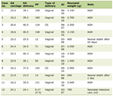
A total of 65 patients were included in the present analysis (male-to-female ratio, 38/27 (58.5%:41.5%), median age 79 (76–83) years). Thirty patients had a PSI class between I and III (79 (75–83) points), while 35 patients were classified as PSI class IV–V (129 (100–156) points). No patient had a PSI class of I, and only four patients were in PSI class II. Among patients with PSI class IV–V, 28 (80%) experienced at least one pneumonia-related complication, and 10 patients (28.6%) died during hospitalization, including eight deaths attributable to CAP.
As expected, patients with PSI class IV–V had higher CRP (P = 0.019), urea (P < 0.001), and creatinine (P < 0.001) levels compared with those in PSI classes I–III, whereas albumin (P = 0.001) and eGFR (P < 0.001) values were lower. Similarly, patients in higher PSI classes had a longer hospital stay (P < 0.001) and a prolonged time to clinical stability (P = 0.002).
The clinical and biochemical characteristics of the study population are summarized in Table 1.
 Click to view | Table 1. Characteristics and Demographics of the Patients |
NP-I and adverse outcomes
NP-I levels across individual PSI categories and aggregated low-intermediate versus high-risk classes are shown in Table 1 and Figure 1. No significant difference was observed between low-intermediate and high-risk groups (242 (138–526) vs. 307 (181–597) pM, P = 0.441). However, when the comparison was restricted to patients in PSI class III versus class V, a trend was noted (183 (130–440) vs. 456 (218–622) pM, P = 0.080).
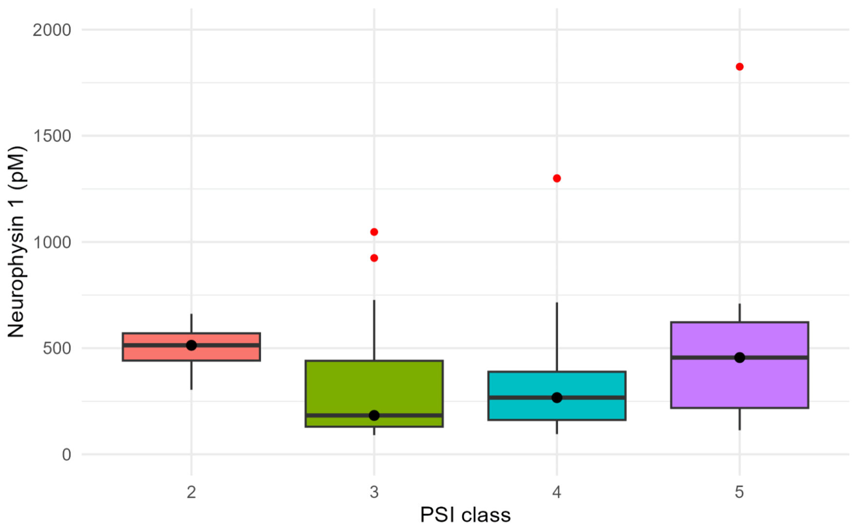 Click for large image | Figure 1. Distribution of Neurophysin I levels across the different PSI risk classes. Three outliers (two in PSI class III and one in PSI class V) were not displayed for visualization purposes. PSI: Pneumonia Severity Index. |
NP-I levels were positively correlated with CRP (R = 0.249, P = 0.045) and inversely correlated with eGFR (R = −0.367, P = 0.003). Patients with impaired kidney function (n = 28, 43.1%) had significantly higher NP-I levels than those with preserved renal function (n = 37, 56.9%; 446 (278–685) vs. 182 (124–403) pM, P < 0.001). In multivariable linear regression adjusted for age and sex, NP-I levels were negatively associated with eGFR (β = −9.060, 95% CI, −17.596 to −0.525, P = 0.038) (Table 2).
Click to view | Table 2. Multivariable Linear Regression Analysis of the Association Between Renal Function and NP-I Concentrations |
No difference in NP-I concentrations was found between patients with and without CAP-related complications (336 (190–607) vs. 225 (147–539) pM, P = 0.326), and this was confirmed in logistic regression analysis for CAP-related complications (OR, 1.000; 95% CI, 0.999–1.005; P = 0.794).
Similarly, NP-I levels did not differ between patients with and without CAP-related death (199 (121–429) vs. 304 (174–604) pM; P = 0.259), and logistic regression confirmed the absence of association with CAP-related mortality (OR, 0.999; 95% CI, 0.995–1.000; P = 0.373).
In univariable logistic regression, PSI points were significantly associated with in-hospital mortality (OR, 1.027; 95% CI 1.008–1.049; P = 0.006), with an AUC of 0.803. Adding NP-I to the model did not significantly improve prediction: PSI remained significant (OR, 1.035; 95% CI, 1.013–1.062; P = 0.004), whereas NP-I was not (OR, 0.997; 95% CI, 0.991–1.000; P = 0.137), and the combined model showed an AUC of 0.829 (DeLong’s test, P = 0.509, compared to PSI alone).
| Discussion | ▴Top |
The results of our study highlight two main findings. First, NP-I measured shortly after CAP diagnosis does not appear to provide additional prognostic information beyond the PSI score alone. Second, NP-I levels are inversely associated with renal function. Indeed, patients with renal impairment exhibited significantly higher concentrations than those with normal kidney function.
In the last years, OXT has increasingly been recognized as a pleiotropic neurohormone involved in reproduction, stress regulation, cardiovascular function and inflammatory processes [25, 26]. However, most of the evidence supporting these actions, including its putative anti-inflammatory effects, derives from animal studies, while robust human data remain limited [26]. Moreover, the majority of existing studies have focused on ischemic and oncologic contexts [11, 12, 27–30], whereas studies addressing inflammation secondary to infection in clinical settings are scarce.
The growing interest in OXT has simultaneously highlighted the substantial methodological challenges associated with its quantification. OXT is a very small peptide (only nine amino acids) and is highly unstable, making accurate measurement technically demanding. Traditional assays such as radioimmunoassays and enzyme immunoassays are hampered by cross-reactivity, limited sensitivity, and suboptimal specificity. Conversely, liquid chromatography–mass spectrometry provides superior analytical performance but is costly, labor-intensive, and not widely available, underscoring the need for more practical alternatives [31–33].
Unlike OXT, its cleavage product NP-I, which is co-released into the circulation in equimolar amounts, is a much larger peptide (94 amino acids). This structural characteristic enables accurate quantification using sandwich immunoassays with paired capture and detection antibodies targeting non-overlapping epitopes. NP-I measurement provides major analytical advantages: it does not require extraction procedures, and total processing time is under 2 h, compared with the 16–24 h typically required for extraction-based OXT assays [16, 17, 34].
In our study, no significant differences in NP-I levels were observed at the time of inclusion across the different PSI risk classes, nor was there any association between NP-I concentrations and the subsequent occurrence of CAP-related complications or in-hospital mortality. Our results confirmed the strong predictive capacity of the PSI score for mortality in hospitalized patients; however, NP-I did not provide additional prognostic value, either alone or in combination with the PSI score. While previous studies have primarily evaluated the impact of exogenous OXT administration on inflammatory responses, evidence regarding circulating OXT or its surrogate markers as predictors of clinical outcomes remains limited.
These findings are consistent with the broader CAP biomarker literature. CRP and PCT reflect the systemic inflammatory response and the host response to infection, but their incremental prognostic value beyond established severity scores has been inconsistent. In a cohort of 925 patients with CAP, admission CRP, PCT, and white blood cell counts did not significantly improve CURB-65-based mortality prediction, whereas follow-up concentrations on days 3–7 provided greater prognostic information [35]. Similarly, a systematic review and meta-analysis concluded that biomarkers predicted short-term mortality with moderate-to-good accuracy but had no clear advantage over CAP-specific clinical scores [36]. Biomarkers reflecting endothelial dysfunction or neuroendocrine stress have shown stronger prognostic associations. In the ProHOSP cohort, adding midregional proadrenomedullin (MR-proADM) improved the predictive performance of both the PSI and CURB-65 scores [37]. Copeptin, a stable surrogate marker of vasopressin, has also been associated with CAP severity and mortality [38] and represents a relevant comparator because, like NP-I, it reflects posterior pituitary secretion. In contrast, NP-I was not associated with adverse outcomes in our cohort. Several factors may explain the absence of a prognostic association. First, NP-I was measured only once, at the time of hospital admission. This might not adequately capture the dynamics of the neuroendocrine response to acute infection. It is conceivable that changes in NP-I levels over time, rather than a single baseline measurement at admission, are more closely related to disease outcome or progression. Second, the pathophysiology of the inflammatory process is highly heterogeneous, influenced by pathogen, immune response, comorbidities and age. Finally, the relatively small sample size and particularly the low number of deaths may have limited our ability to detect an association between NP-I and clinical outcomes or to demonstrate incremental prognostic value beyond an established multidimensional score such as the PSI. A weak positive correlation was observed between NP-I and CRP. Preclinical studies in mice have shown that OXT administration can reduce CRP levels, suggesting that the modest correlation in our cohort could reflect a compensatory increase of the oxytocinergic system in response to rising inflammation rather than a direct anti-inflammatory effect detectable at the systemic level [25]. Interestingly, OXT has been shown to buffer neuroendocrine stress responses in different settings. However, given the weak correlation, its clinical relevance for inflammation control or outcomes in patients with CAP remains uncertain.
Notably, any potential association between NP-I and inflammatory burden might be confounded by renal function, which appears to be an important determinant of circulating NP-I concentrations. Our results revealed a significant association between renal function and NP-I levels, an observation that was also confirmed in the multivariable regression model. Human data on NP-I metabolism are scarce. Early animal studies by Morris et al in the 1970s demonstrated a key role of the kidney in neurophysin clearance, with nephrectomized rats exhibiting a marked prolongation of circulating half-life [39]. Our findings are consistent with these experimental observations and suggest that reduced renal elimination may contribute to the higher circulating NP-I concentrations observed in patients with reduced eGFR.
Dependence on renal function is also recognized for other peptide biomarkers. Copeptin concentrations increase as renal function declines, and its correspondence with vasopressin becomes less reliable in advanced renal impairment [40]. Reduced eGFR also affects concentrations of natriuretic peptides, PCT and MR-proADM [41–43], complicating their interpretation as markers of secretion or disease activity. NP-I may be subject to a similar limitation. Elevated NP-I concentrations in patients with reduced eGFR should therefore not automatically be interpreted as evidence of increased oxytocinergic activity. Overall, although NP-I did not improve prognostic assessment in CAP, our findings provide novel clinical information on the interpretation of circulating NP-I concentrations and highlight renal function as an important factor to consider when evaluating NP-I as a biomarker in future studies.
Our study has several strengths and limitations. Strengths include the prospective design of the original cohort, with patients followed from the time of CAP diagnosis in the emergency department or medical wards, and the evaluation of a novel biomarker for which clinical data in infection-related settings are currently limited.
Limitations of our study include the relatively small sample size and the low number of CAP-related deaths. Moreover, patients in PSI class I were not represented, and those in class II were underrepresented, so we cannot determine whether including these lower-risk categories might have revealed a greater discriminative ability of NP-I for adverse outcomes. NP-I was measured only at the time of patient inclusion; therefore, we cannot assess whether longitudinal changes, rather than a single baseline value, might provide additional prognostic information. In addition, data on proteinuria, albuminuria, and smoking status were not available, precluding assessment of their potential influence on NP-I concentrations. Finally, as a secondary post hoc analysis, the possibility of selection bias cannot be entirely excluded. Importantly, only patients with normal serum sodium were included to reduce potential confounding, as the impact of hyponatremia on OXT or NP-I levels is not well established. While this approach improved cohort homogeneity, it may limit the generalizability of our findings to patients with dysnatremia.
Conclusions
In conclusion, our study shows that NP-I measured shortly after the diagnosis of CAP does not appear to have the ability to predict adverse outcomes, such as CAP-related complications or death. Indeed, the addition of NP-I to the PSI score did not provide clinicians with any meaningful improvement in risk stratification.
NP-I levels were significantly higher in patients with impaired renal function, which may reflect reduced renal clearance and its consequent accumulation in circulation.
Future prospective studies are needed to determine whether NP-I might potentially provide prognostic information to distinguish patients in lower PSI classes (I–II)—which were underrepresented in our cohort—from those at higher risk, and to further explore its potential prognostic relevance in other infectious diseases beyond pulmonary involvement.
Acknowledgments
We thank the staff of the emergency departments, medical wards, and laboratories of all participating hospitals for their support of this study. We also acknowledge the collaborators, as well as the study and laboratory personnel across all participating sites, whose contributions made this trial possible. Finally, we are deeply grateful to all patients for their participation.
Financial Disclosure
This study was supported by a grant from the Swiss National Foundation (PP0P3_123346) to MC-C.
Conflict of Interest
MEL is a consultant for Mind Medicine Inc. and Lykos Therapeutics. All other authors declare no competing interests.
Informed Consent
Written informed consent was obtained from all participants before study enrollment.
Author Contributions
EV contributed to data collection, data analysis, data interpretation, and the literature search, and wrote and edited the manuscript. AN contributed to data collection, data analysis, data interpretation, and the literature search, and wrote and edited the manuscript. DR, DL and MEL performed the measurements and edited the manuscript. CAB, NC-N, BM and MC-C designed the original study and wrote the protocol. CAB, NC-N, MB, PS, NR, BM and MC-C contributed to data interpretation and edited the manuscript.
Data Availability
The data sets generated during and/or analyzed during the current study are not publicly available but are available from the corresponding author on reasonable request.
Abbreviations
AUC: area under the curve; BMI: body mass index; CAP: community-acquired pneumonia; CI: confidence interval; CKD-EPI: Chronic Kidney Disease Epidemiology Collaboration; CRP: C-reactive protein; CV: coefficient of variation; eGFR: estimated glomerular filtration rate; IQR: interquartile range; NP-I: neurophysin I; OR: odds ratio; OXT: oxytocin; PCT: procalcitonin; PSI: Pneumonia Severity Index; ROC: receiver operating characteristic
| References | ▴Top |
- The top 10 causes of death (Internet). cited Nov 18, 2025). https://www.who.int/news-room/fact-sheets/detail/the-top-10-causes-of-death. Accessed Nov 18, 2025.
- Vaughn VM, Dickson RP, Horowitz JK, Flanders SA. Community-acquired pneumonia: a review. JAMA. 2024;332(15):1282-1295.
doi pubmed - Regunath H, Oba Y. Community-acquired pneumonia. StatPearls (Internet). Treasure Island (FL): StatPearls Publishing; 2025 (cited Apr 14, 2025). http://www.ncbi.nlm.nih.gov/books/NBK430749/. Accessed Apr 14, 2025.
- Christ-Crain M, Jaccard-Stolz D, Bingisser R, Gencay MM, Huber PR, Tamm M, Muller B. Effect of procalcitonin-guided treatment on antibiotic use and outcome in lower respiratory tract infections: cluster-randomised, single-blinded intervention trial. Lancet. 2004;363(9409):600-607.
doi pubmed - Schuetz P, Wirz Y, Sager R, Christ-Crain M, Stolz D, Tamm M, Bouadma L, et al. Procalcitonin to initiate or discontinue antibiotics in acute respiratory tract infections. Cochrane Database Syst Rev. 2017;10(10):CD007498.
doi pubmed - Varaldo E, Rumbolo F, Prencipe N, Bioletto F, Settanni F, Mengozzi G, Grottoli S, et al. Effectiveness of Copeptin, MR-proADM and MR-proANP in predicting adverse outcomes, alone and in combination with traditional severity scores, a secondary analysis in COVID-19 patients requiring intensive care admission. J Clin Med. 2024;13(7):2019.
doi pubmed - Karakioulaki M, Stolz D. Biomarkers in pneumonia-beyond procalcitonin. Int J Mol Sci. 2019;20(8):2004.
doi pubmed - Uvnas-Moberg K, Gross MM, Calleja-Agius J, Turner JD. The Yin and Yang of the oxytocin and stress systems: opposites, yet interdependent and intertwined determinants of lifelong health trajectories. Front Endocrinol (Lausanne). 2024;15:1272270.
doi pubmed - Olff M, Frijling JL, Kubzansky LD, Bradley B, Ellenbogen MA, Cardoso C, Bartz JA, et al. The role of oxytocin in social bonding, stress regulation and mental health: an update on the moderating effects of context and interindividual differences. Psychoneuroendocrinology. 2013;38(9):1883-1894.
doi pubmed - Carter CS. Oxytocin and love: Myths, metaphors and mysteries. Compr Psychoneuroendocrinol. 2022;9:100107.
doi pubmed - Momenabadi S, Vafaei AA, Bandegi AR, Zahedi-Khorasani M, Mazaheri Z, Vakili A. Oxytocin reduces brain injury and maintains blood-brain barrier integrity after ischemic stroke in mice. Neuromolecular Med. 2020;22(4):557-571.
doi pubmed - Yang M, Deng S, Jiang J, Tian M, Xiao L, Gong Y. Oxytocin improves intracerebral hemorrhage outcomes by suppressing neuronal pyroptosis and mitochondrial fission. Stroke. 2023;54(7):1888-1900.
doi pubmed - Christ-Crain M, Stolz D, Jutla S, Couppis O, Muller C, Bingisser R, Schuetz P, et al. Free and total cortisol levels as predictors of severity and outcome in community-acquired pneumonia. Am J Respir Crit Care Med. 2007;176(9):913-920.
doi pubmed - Kellum JA, Kong L, Fink MP, Weissfeld LA, Yealy DM, Pinsky MR, Fine J, et al. Understanding the inflammatory cytokine response in pneumonia and sepsis: results of the Genetic and Inflammatory Markers of Sepsis (GenIMS) Study. Arch Intern Med. 2007;167(15):1655-1663.
doi pubmed - Gimpl G, Fahrenholz F. The oxytocin receptor system: structure, function, and regulation. Physiol Rev. 2001;81(2):629-683.
doi pubmed - MacLean EL, Carranza E, Gnanadesikan GE, King KM, Allen AM, Linde-Krieger LB, Feldman R, et al. Neurophysin I is an analytically robust surrogate biomarker for oxytocin. Psychoneuroendocrinology. 2024;161:106951.
doi pubmed - Atila C, Nikaj A, Leibnitz S, Liechti ME, Christ-Crain M. Neurophysin I: a reliable, novel, and robust biomarker for oxytocin. Eur J Endocrinol. 2025;192(4):502-510.
doi pubmed - Amico JA, Seif SM, Robinson AG. Oxytocin in human plasma: correlation with neurophysin and stimulation with estrogen. J Clin Endocrinol Metab. 1981;52(5):988-993.
doi pubmed - Tabak BA, Leng G, Szeto A, Parker KJ, Verbalis JG, Ziegler TE, Lee MR, et al. Advances in human oxytocin measurement: challenges and proposed solutions. Mol Psychiatry. 2023;28(1):127-140.
doi pubmed - Leibnitz S, Christ-Crain M, Atila C. Oxytocin: a neglected hormone in pituitary disease - From function to the diagnosis of a deficiency, resulting clinical relevance, and potential treatment options in endocrinology. Arch Endocrinol Metab. 2025;70(special 1):e20250259.
doi pubmed - Blum CA, Nigro N, Briel M, Schuetz P, Ullmer E, Suter-Widmer I, Winzeler B, et al. Adjunct prednisone therapy for patients with community-acquired pneumonia: a multicentre, double-blind, randomised, placebo-controlled trial. Lancet. 2015;385(9977):1511-1518.
doi pubmed - Fine MJ, Auble TE, Yealy DM, Hanusa BH, Weissfeld LA, Singer DE, Coley CM, et al. A prediction rule to identify low-risk patients with community-acquired pneumonia. N Engl J Med. 1997;336(4):243-250.
doi pubmed - Nikaj A, Atila C, Rudin D, Luethi D, Sailer CO, Winzeler B, Urwyler SA, et al. Neurophysin-I dynamics upon different pituitary provocation tests in healthy participants. Endocr Connect. 2026;15(5):e250929.
doi pubmed - R Core Team. R: A language and environment for statistical computing. R Foundation for Statistical Computing. https://www.R-project.org/. 2024.
- Mehdi SF, Pusapati S, Khenhrani RR, Farooqi MS, Sarwar S, Alnasarat A, Mathur N, et al. Oxytocin and Related Peptide Hormones: Candidate Anti-Inflammatory Therapy in Early Stages of Sepsis. Front Immunol. 2022;13:864007.
doi pubmed - Aulinas A, Lawson EA. The Oxytocin System and Implications for Oxytocin Deficiency in Hypothalamic-Pituitary Disease. Endocr Rev. 2025;46(4):518-548.
doi pubmed - Jankowski M, Bissonauth V, Gao L, Gangal M, Wang D, Danalache B, Wang Y, et al. Anti-inflammatory effect of oxytocin in rat myocardial infarction. Basic Res Cardiol. 2010;105(2):205-218.
doi pubmed - Buemann B, Uvnas-Moberg K. Oxytocin may have a therapeutical potential against cardiovascular disease. Possible pharmaceutical and behavioral approaches. Med Hypotheses. 2020;138:109597.
doi pubmed - Nation DA, Szeto A, Mendez AJ, Brooks LG, Zaias J, Herderick EE, Gonzales J, et al. Oxytocin attenuates atherosclerosis and adipose tissue inflammation in socially isolated ApoE-/- mice. Psychosom Med. 2010;72(4):376-382.
doi pubmed - Cuneo MG, Szeto A, Schrepf A, Kinner EM, Schachner BI, Ahmed R, Thaker PH, et al. Oxytocin in the tumor microenvironment is associated with lower inflammation and longer survival in advanced epithelial ovarian cancer patients. Psychoneuroendocrinology. 2019;106:244-251.
doi pubmed - MacLean EL, Wilson SR, Martin WL, Davis JM, Nazarloo HP, Carter CS. Challenges for measuring oxytocin: The blind men and the elephant? Psychoneuroendocrinology. 2019;107:225-231.
doi pubmed - Franke AA, Li X, Dabalos C, Lai JF. Improved oxytocin analysis from human serum and urine by orbitrap ESI-LC-HRAM-MS. Drug Test Anal. 2020;12(6):846-852.
doi pubmed - Franke AA, Li X, Menden A, Lee MR, Lai JF. Oxytocin analysis from human serum, urine, and saliva by orbitrap liquid chromatography-mass spectrometry. Drug Test Anal. 2019;11(1):119-128.
doi pubmed - Szeto A, McCabe PM, Nation DA, Tabak BA, Rossetti MA, McCullough ME, Schneiderman N, et al. Evaluation of enzyme immunoassay and radioimmunoassay methods for the measurement of plasma oxytocin. Psychosom Med. 2011;73(5):393-400.
doi pubmed - Zhydkov A, Christ-Crain M, Thomann R, Hoess C, Henzen C, Werner Z, Mueller B, et al. Utility of procalcitonin, C-reactive protein and white blood cells alone and in combination for the prediction of clinical outcomes in community-acquired pneumonia. Clin Chem Lab Med. 2015;53(4):559-566.
doi pubmed - Viasus D, Del Rio-Pertuz G, Simonetti AF, Garcia-Vidal C, Acosta-Reyes J, Garavito A, Carratala J. Biomarkers for predicting short-term mortality in community-acquired pneumonia: a systematic review and meta-analysis. J Infect. 2016;72(3):273-282.
doi pubmed - Schuetz P, Wolbers M, Christ-Crain M, Thomann R, Falconnier C, Widmer I, Neidert S, et al. Prohormones for prediction of adverse medical outcome in community-acquired pneumonia and lower respiratory tract infections. Crit Care. 2010;14(3):R106.
doi pubmed - Kruger S, Ewig S, Kunde J, Hartmann O, Suttorp N, Welte T, CAPNETZ Study Group. Pro-atrial natriuretic peptide and pro-vasopressin for predicting short-term and long-term survival in community-acquired pneumonia: results from the German Competence Network CAPNETZ. Thorax. 2010;65(3):208-214.
doi pubmed - Morris BJ, Hutchinson JS, Johnston CI. Clearance and metabolism of neurophysins by rat kidney. J Endocrinol. 1975;64(2):201-213.
doi pubmed - Ettema EM, Heida J, Casteleijn NF, Boesten L, Westerhuis R, Gaillard C, Gansevoort RT, et al. The effect of renal function and hemodialysis treatment on plasma vasopressin and copeptin levels. Kidney Int Rep. 2017;2(3):410-419.
doi pubmed - Takase H, Dohi Y. Kidney function crucially affects B-type natriuretic peptide (BNP), N-terminal proBNP and their relationship. Eur J Clin Invest. 2014;44(3):303-308.
doi pubmed - Wu SC, Liang CX, Zhang YL, Hu WP. Elevated serum procalcitonin level in patients with chronic kidney disease without infection: a case-control study. J Clin Lab Anal. 2020;34(2):e23065.
doi pubmed - Krintus M, Kozinski M, Braga F, Kubica J, Sypniewska G, Panteghini M. Plasma midregional proadrenomedullin (MR-proADM) concentrations and their biological determinants in a reference population. Clin Chem Lab Med. 2018;56(7):1161-1168.
doi pubmed
This article is distributed under the terms of the Creative Commons Attribution 4.0 International License (CC BY 4.0), which permits unrestricted use, distribution, and reproduction in any medium, including commercial use, provided the original work is properly cited.
Journal of Clinical Medicine Research is published by Elmer Press Inc.