| Journal of Clinical Medicine Research, ISSN 1918-3003 print, 1918-3011 online, Open Access |
| Article copyright, the authors; Journal compilation copyright, J Clin Med Res and Elmer Press Inc |
| Journal website https://jocmr.elmerjournals.com |
Original Article
Volume 18, Number 11, November 2026, pages 823-834

Serum Serotonin and Noninvasive Portal Hypertension Indices in Relation to Esophageal Variceal Severity in Cirrhosis: An Exploratory Cross-Sectional Study
Amir M. Farida, r, Ahmed Moustafa ElFayoumyb, c, r, Awatif Elmohamady Edreesd, e, Fatma Saffeyeldin Mohamede, f, Saifeliazal Abdelazim Suliman Abdelwahidg, Nagwa Roshdy Mohamedh, i, Osama Mostafa Abd Elrazikj, Ahmed Akefk, s, Ashraf Ahmed Mohamed Aboismaill, Ahmed Abdelmalikj, Marina Albert Megallym, Ahmed Arakiepn, Abdellatif Salah Eldin Elolimyo, Inas Abdullah Mohamed Issa Barakatp, Saeed M. El-Nahaasq
aDepartment of Tropical Medicine, Faculty of Medicine, Ain Shams University, Cairo, Egypt
bDepartment of Gastroenterology, Faculty of Medicine, Ain Shams University, Cairo, Egypt
cDepartment of Gastroenterology, Cumberland Infirmary, North Cumbria Integrated Care NHS Foundation Trust, Carlisle, UK
dDepartment of Tropical Medicine, Faculty of Medicine, Tanta University, Tanta, Egypt
eDepartment of Internal Medicine, College of Medicine, Taif University, Taif, Saudi Arabia
fDepartment of Hepatogastroenterology and Infectious Diseases, Faculty of Medicine for Girls, Al-Azhar University, Cairo, Egypt
gDepartment of Clinical Excellence, Bupa Arabia, Jeddah, Saudi Arabia
hDepartment of Internal Medicine, Faculty of Medicine, Ain Shams University, Cairo, Egypt
iInternal Medicine and Endocrinology, Mouwasat Hospital, Jubail, Saudi Arabia
jDepartment of Hepatology, Gastroenterology and Infectious Diseases, Faculty of Medicine, Al-Azhar University, Cairo, Egypt
kDepartment of Clinical Pathology, Faculty of Medicine, Mansoura University, Mansoura, Egypt
lDepartment of Tropical Medicine, Faculty of Medicine, Alexandria University, Alexandria, Egypt
mDepartment of Internal Medicine, Faculty of Medicine, Alexandria University, Alexandria, Egypt
nEmergency Department, Faculty of Medicine, Tanta University, Tanta, Egypt
oDepartment of Radiodiagnosis and Interventional Radiology, National Liver Institute, Menoufia University, Shebin El-Kom, Egypt
pDepartment of Internal Medicine, Taibah University, Medina, Saudi Arabia
qEndemic Medicine Department and Hepatology Unit, Faculty of Medicine, Cairo University, Cairo, Egypt
rThese authors contributed equally to this work.
sCorresponding Author: Ahmed Akef, Department of Clinical Pathology, Faculty of Medicine, Mansoura University, Mansoura 35516, Egypt
Manuscript submitted September 30, 2026, accepted October 9, 2026, published online November 9, 2026
Short title: Serotonin and Noninvasive Indices in EV Severity
doi: https://doi.org/10.14740/jocmr6742
| Abstract | ▴Top |
Background: Esophageal varices are a life-threatening consequence of portal hypertension in liver cirrhosis, and upper gastrointestinal endoscopy, the reference standard for detecting them, is invasive and resource-intensive. Serum serotonin, over 90% of which is stored in circulating platelets, has been proposed as a candidate biochemical correlate of portal hypertensive and fibrotic change, but its relationship to variceal severity alongside established noninvasive indices has not been well characterized. This study aimed to determine the association of serum serotonin, Child-Pugh class, portal vein diameter, and platelet count to spleen diameter ratio (PSR) with endoscopically graded esophageal variceal severity in patients with liver cirrhosis, and to explore the diagnostic performance of these markers for identifying high-grade varices.
Methods: In this exploratory cross-sectional study, 140 participants were enrolled into five prespecified groups of fixed size: non-hepatic controls (n = 40), cirrhosis without varices (n = 40), and grade 1 (n = 20), grade 2 (n = 20), and grade 3 (n = 20) esophageal varices confirmed endoscopically. Serum serotonin was measured by enzyme-linked immunosorbent assay (ELISA). Group comparisons used one-way ANOVA or the Kruskal-Wallis test according to the normality of model residuals, and the chi-square or Fisher-Freeman-Halton exact test for categorical variables. Spearman correlation and multivariable ordinal logistic regression identified independent predictors of variceal grade among the 60 participants with varices, and receiver operating characteristic (ROC) analysis assessed diagnostic performance for grade 2/3 varices among the 99 cirrhotic participants with complete data, with bootstrap correction of cutoff-based estimates for optimism and pairwise DeLong comparison of AUCs; a Firth-penalized model was fitted as a sensitivity analysis.
Results: Serum serotonin rose progressively across groups, from 30.0 ± 5.6 ng/mL in controls to 106.5 ± 5.2 ng/mL in grade 3 varices (F = 347.575, P < 0.001), and was, together with portal vein diameter, an independent predictor of variceal grade after adjustment (adjusted odds ratio (OR) of 7.48 per 10 ng/mL, P = 0.0009; portal vein diameter adjusted OR of 1.70 per 0.1 mm, P = 0.0002; exploratory model, n = 59). Apparent areas under the curve (AUCs) for grade 2/3 varices were 0.944 (95% confidence interval (CI), 0.895–0.980) for serotonin, 0.923 (0.864–0.974) for portal vein diameter, 0.764 for PSR, and 0.746 for Child-Pugh class; serotonin and portal vein diameter did not differ significantly (DeLong P = 0.553), whereas each exceeded PSR and Child-Pugh class (all P ≤ 0.004). Both associations persisted in the penalized model, and optimism correction changed the accuracy estimates only slightly.
Conclusions: In this exploratory study, serum serotonin tracked with esophageal variceal severity and, alongside portal vein diameter, was independently associated with variceal grade, with discrimination comparable to that of portal vein diameter. Because the sample was enriched by design and cutoffs were derived and tested in the same cohort, these findings are hypothesis-generating and require validation in unselected surveillance cohorts before clinical use.
Keywords: Liver cirrhosis; Esophageal and gastric varices; Serotonin; Child-Pugh score; Portal hypertension; Platelet count to spleen diameter ratio
| Introduction | ▴Top |
Liver cirrhosis is the common histological endpoint of chronic liver injury and remains a major contributor to global mortality, with cirrhosis-related deaths continuing to rise across most regions over recent decades [1, 2]. Portal hypertension, the principal hemodynamic consequence, drives the formation of portosystemic collaterals, of which esophageal varices are the most clinically important. Once formed, varices can enlarge over time and eventually rupture, and variceal hemorrhage remains one of the leading causes of death in decompensated cirrhosis, carrying substantial short-term mortality despite advances in endoscopic and pharmacological therapy over the past two decades. Roughly half of patients already have esophageal varices at the time cirrhosis is first diagnosed, and this proportion rises further as hepatic decompensation progresses, making reliable identification of at-risk patients a recurring surveillance need rather than a single assessment at diagnosis.
Upper gastrointestinal endoscopy remains the reference standard for detecting and grading esophageal varices, and current international consensus continues to recommend endoscopic assessment in patients at elevated risk of variceal hemorrhage [3]. Endoscopy is nonetheless invasive, requires sedation and dedicated resources, and is poorly tolerated by some patients, particularly where repeated surveillance is required over many years. These practical limitations have sustained a long-standing effort to identify noninvasive clinical, biochemical, and ultrasonographic parameters capable of triaging patients toward, or safely away from, endoscopic assessment.
The Child-Pugh classification, derived from serum bilirubin, albumin, prothrombin time, ascites, and hepatic encephalopathy, was originally developed to stratify surgical risk in cirrhosis and remains one of the most widely used prognostic tools in chronic liver disease [4, 5]; it correlates broadly with the severity of portal hypertensive complications, although it reflects hepatic synthetic reserve rather than variceal anatomy directly. Portal vein diameter, measured by abdominal ultrasound, offers a simple surrogate of portal hypertensive hemodynamics that has been associated with the presence of varices [6], although its measurement is influenced by respiratory phase, hydration status, and operator technique. Thrombocytopenia secondary to hypersplenism is a further downstream consequence of portal hypertension, and the platelet count to spleen diameter ratio (PSR), which combines both effects into a single index, has been evaluated across several cohorts as a correlate of variceal presence and severity [7–9], although its diagnostic accuracy varies with the population studied and the cutoff selected. None of these three indices was developed with the others in mind, and although each has been validated individually, their relative and combined performance when measured together in the same patients, alongside a candidate biochemical marker, has rarely been reported.
Serotonin (5-hydroxytryptamine) is synthesized largely outside the liver but is taken up and stored almost entirely within circulating platelets, from which it is released on platelet activation to influence vascular tone, hepatic microcirculation, and liver regeneration [10, 11]. Platelet-derived serotonin modulates hepatic stellate cell activation and fibrogenic signaling [12], and blockade of hepatic serotonin-receptor signaling has attenuated fibrosis in experimental models [13], raising the possibility that circulating serotonin concentration tracks the severity of the portal hypertensive and fibrotic processes that underlie variceal formation. Whether serum serotonin adds discriminative information beyond Child-Pugh class, portal vein diameter, and PSR, rather than simply mirroring them, has not been well characterized in a single cohort.
Acute variceal hemorrhage is also an important precipitant of hepatic encephalopathy, a complication that can require urgent neurological and airway assessment in patients with advanced cirrhosis [14], which reinforces the practical value of identifying patients with high-grade varices before bleeding occurs rather than after. This study therefore examined the association of serum serotonin, Child-Pugh class, portal vein diameter, and PSR with endoscopically graded esophageal variceal severity in a cohort spanning non-hepatic controls, cirrhosis without varices, and grade 1 to grade 3 varices, and explored the diagnostic performance of these four markers, individually, for identifying high-grade varices. The diagnostic analyses were designed as exploratory and hypothesis-generating, not as a validation of any marker or cutoff.
| Materials and Methods | ▴Top |
Study design, setting, and ethical considerations
This study was conducted in accordance with the ethical principles of the Declaration of Helsinki [15] and with the ethical standards of the responsible institution on human subjects and is reported in accordance with the Strengthening the Reporting of Observational Studies in Epidemiology (STROBE) guideline for observational studies [16]. Ethical approval was obtained from the Institutional Review Board (IRB) of the Faculty of Medicine, Al-Azhar University, Cairo, Egypt (IRB: AZFM-IRB/2026/07/0087) prior to data collection, and written informed consent was obtained from each participant before enrollment. Participant identifiers were replaced with study codes before analysis, and confidentiality was maintained throughout data handling and reporting.
Study population
A total of 140 adults were enrolled: patients with an established diagnosis of liver cirrhosis undergoing upper gastrointestinal endoscopy, and age- and sex-comparable adults without liver disease who underwent upper gastrointestinal endoscopy for other indications and served as non-hepatic controls. Participants were assigned to one of five groups on the basis of clinical status and endoscopic findings: non-hepatic controls (n = 40); cirrhosis without esophageal varices (n = 40); and cirrhosis with grade 1 (n = 20), grade 2 (n = 20), or grade 3 (n = 20) esophageal varices, graded according to a standard three-tier endoscopic classification of variceal size [17]. Participants were excluded if they had non-cirrhotic portal hypertension, a prior portosystemic shunt or definitive variceal eradication procedure, a hematological disorder independently causing thrombocytopenia or splenomegaly, or incomplete data for the primary predictors or the outcome. Patients with hepatocellular carcinoma or another gastrointestinal malignancy, portal or splenic vein thrombosis, neuropsychiatric disorders, or known carcinoid syndrome, and those taking nonsteroidal anti-inflammatory drugs or selective serotonin reuptake inhibitors, were also excluded because these conditions and drugs can alter circulating serotonin or portal hemodynamics. This five-group design was chosen deliberately so that each candidate marker could be evaluated across the full spectrum of disease, from the absence of cirrhosis, through cirrhosis without portal hypertensive complications, to progressively more severe varices, rather than only across the narrower range of a single cirrhotic population. Non-hepatic controls were included to characterize the range of serum serotonin in the absence of liver disease and were excluded from all regression and diagnostic analyses, which were restricted to participants with cirrhosis.
Participants were recruited from adults (older than 18 years) presenting for upper gastrointestinal endoscopy to the endoscopy units of the Department of Hepatology, Gastroenterology and Infectious Diseases, Al-Azhar University Hospitals, Cairo, Egypt. Eligible patients were enrolled and, after endoscopy, allocated to the corresponding stratum; enrollment into each stratum closed once its prespecified target size (40 without varices, 20 for each variceal grade, and 40 non-hepatic controls) had been reached. Because this stratified (quota) design fixed the number of participants in each variceal grade, the case mix differs from that of an unselected population undergoing variceal surveillance, in which absent or small varices predominate; this was taken into account in the analysis and interpretation of diagnostic performance. Serum serotonin data from this cohort were previously reported in part [18], in an analysis of serotonin in relation to the presence of varices; the present study addresses different questions, namely the joint evaluation of serotonin with Child-Pugh class, portal vein diameter, and PSR, multivariable ordinal modeling, and paired ROC comparison, using the verified participant-level dataset.
Clinical, laboratory, and serotonin assessment
Demographic and clinical data, including duration of liver disease, jaundice, ascites, gastrointestinal bleeding, peripheral edema, and clinically detected splenomegaly, were extracted using a standardized form. Platelet count, prothrombin time, international normalized ratio (INR), total and direct bilirubin, alanine aminotransferase (ALT) and aspartate aminotransferase (AST), alkaline phosphatase, albumin, and total protein were measured using standard laboratory methods. Serum serotonin concentration was measured using a Human Serotonin enzyme-linked immunosorbent assay (ELISA) kit (Catalogue No. E1128Hu; BT LAB/Bioassay Technology Laboratory, Shanghai, China) according to the manufacturer’s instructions, with optical density read at 450 nm and concentration derived from the kit’s calibration curve. Because circulating serotonin is closely tied to platelet handling, blood collection and processing followed a standardized protocol intended to limit pre-analytical variability from platelet degranulation. A 10-mL venous blood sample was obtained from each participant: 2 mL was collected into ethylenediaminetetraacetic acid for the complete blood count, 2 mL into sodium citrate for coagulation studies, and 6 mL into a plain tube without anticoagulant, which was centrifuged at 4,000 revolutions per minute for 10 min; the separated serum was stored frozen at −20 °C until batch analysis. In the serotonin assay, 40 µL of serum, 10 µL of biotinylated anti-serotonin antibody, and 50 µL of streptavidin-horseradish peroxidase were incubated for 60 min at 37 °C; after five washes, substrate solutions A and B were added, and the mixture was incubated for 10 min at 37 °C in the dark. The reaction was stopped, and optical density was read at 450 nm within 10 min. Concentrations were derived from a standard curve fitted by regression to the kit standards. Assay precision was not re-verified in-house; the manufacturer-stated intra-assay and inter-assay coefficients of variation are < 8% and < 10%, respectively. Because serum was stored for batch analysis, serotonin results were not available to the endoscopists or sonographers at the time of their assessments. Because serum rather than platelet-poor plasma was analyzed, the measured concentration predominantly reflects platelet-stored serotonin released during clotting [10].
Endoscopic assessment
Upper gastrointestinal endoscopy was performed in all participants, including the non-hepatic controls, using a Pentax EG34-i10 video gastroscope (Pentax Medical, Tokyo, Japan), and was the reference standard for the presence and grade of esophageal varices. Varices were graded by size as small (grade 1), medium (grade 2), or large (grade 3) using the standard three-tier classification [17]. Grading was recorded by the performing endoscopist at the time of the procedure, without knowledge of serum serotonin results, which were generated later; an independent second reading of the endoscopic findings was not documented, so inter-observer agreement could not be assessed.
Ultrasonographic assessment and derived indices
Abdominal ultrasound was used to record liver size and echogenicity, portal vein diameter, splenic vein diameter, spleen diameter, and the presence and grade of ascites. Child-Pugh class (A, B, or C) was obtained from the clinical dataset and analyzed as an ordinal variable. Because the individual component scores, including encephalopathy grade, were not available as discrete variables, the numerical Child-Pugh score could not be independently reconstructed and Child-Pugh class was therefore analyzed as an ordinal variable rather than as a continuous score [4, 5]. PSR was calculated for each participant individually as platelet count (× 109/L) divided by spleen diameter (cm), multiplied by 100, which is equivalent to the conventional ratio of platelet count (per mm3) to spleen diameter (mm).
Data verification
Before analysis, the dataset was checked for completeness and internal consistency. One record, from a participant with grade 3 varices, contained a non-numeric entry for serum serotonin and was treated as missing for serotonin-dependent analyses only, without substitution; the complete-case number of participants analyzed is reported alongside each result. One spleen diameter value was recorded with an implausible decimal placement (1,372 cm) and was corrected to the value consistent with that same participant’s independently recorded PSR (13.72 cm); no other values were altered, and no missing data were imputed or calculated in place of an absent measurement anywhere in the dataset. Group-level PSR was reported throughout as the mean of each participant’s individually calculated ratio, since the ratio of the group-mean platelet count to the group-mean spleen diameter is a mathematically distinct quantity and was not used.
Statistical analysis
Continuous variables are presented as mean ± standard deviation (SD) or median (interquartile range (IQR)) according to distribution and were compared across the five study groups using one-way analysis of variance (ANOVA) or the Kruskal–Wallis test; the choice of test for each variable was determined by the Shapiro–Wilk normality of the residuals from a group-wise linear model, rather than applied uniformly across all variables. Categorical variables were presented as n (%) and compared using the chi-square test, or the Fisher-Freeman-Halton exact test where more than 20% of expected cell counts were below 5. Among the 60 participants with grade 1–3 varices, the univariable association between each predictor and ordinal variceal grade was assessed using Spearman rank correlation, and independent predictors were identified using a multivariable ordinal (proportional-odds) logistic regression model [19], with serum serotonin (per 10 ng/mL), Child-Pugh class (coded ordinally as A = 1, B = 2, and C = 3), portal vein diameter (per 0.1 mm, because its range among participants with varices was narrow), PSR (per 100-unit decrease), age, and sex entered as covariates; platelet count and spleen diameter were not entered alongside PSR because of their direct mathematical overlap with it. Because the model estimated six parameters from 59 participants distributed across three outcome categories, which is below commonly recommended sample-size criteria for multivariable modeling [20], it was regarded as exploratory, and no variable selection was performed. To assess the stability of the estimates, the model was refitted by Firth-type penalized maximum likelihood, which reduces small-sample bias and yields finite estimates under quasi-separation [21]; a parsimonious model containing only serum serotonin and portal vein diameter was also fitted, and the full model was refitted in 1,000 bootstrap resamples to examine the consistency of the direction of each association. Diagnostic performance for identifying grade 2 or 3 varices was assessed, as an exploratory analysis, by receiver operating characteristic (ROC) analysis among the 99 cirrhotic participants with a complete serotonin measurement, with cutoffs selected by the Youden index [22] and 95% confidence intervals (CIs) for the area under the curve (AUC) obtained by stratified bootstrap resampling. Because the optimal cutoffs were derived and evaluated in the same sample, apparent sensitivity and specificity were corrected for optimism by bootstrap internal validation (1,000 stratified resamples, re-deriving the Youden cutoff in each resample and applying it to the original sample) [23] and were also estimated by leave-one-out cross-validation; the AUC of the exploratory combined model was corrected for optimism in the same way. External validation was not possible within this study. Ninety-five percent CIs for sensitivity and specificity were calculated by the Wilson score method, and positive and negative likelihood ratios, which do not depend on prevalence, were derived from them. Because predictive values depend on the prevalence of high-grade varices, which was fixed at 39.4% by the stratified design, they were additionally recalculated by Bayes’ theorem at illustrative prevalences of 10% and 25%. The AUCs of the individual markers were compared pairwise using DeLong’s nonparametric test for correlated ROC curves [24]. All statistical tests were two-tailed, and P < 0.05 was considered statistically significant. Analyses were performed in Python (SciPy, statsmodels, and scikit-learn).
| Results | ▴Top |
Baseline demographic and clinical characteristics
Of the 140 participants, 40 were non-hepatic controls, 40 had cirrhosis without varices, and 60 had cirrhosis with grade 1 (n = 20), grade 2 (n = 20), or grade 3 (n = 20) esophageal varices (Table 1). Age did not differ significantly across the five groups (H = 9.120, P = 0.058), and the proportion of male participants was similar across all groups (χ2 = 2.227, P = 0.694). Among the four cirrhotic groups, duration of liver disease was longer with increasing variceal grade (H = 26.589, P < 0.001). Jaundice, gastrointestinal bleeding, peripheral edema, and clinically detected splenomegaly were each more frequent with higher variceal grade (all P ≤ 0.002), while ascites showed a similar but nonsignificant trend across groups (Fisher–Freeman–Halton exact P = 0.068). No participant was excluded after enrollment, so the five group sizes reported here (40, 40, 20, 20, and 20) represent the full analyzed cohort for every baseline variable in Table 1.
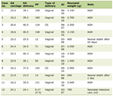 Click to view | Table 1. Baseline Demographic and Clinical Characteristics of the Study Cohort |
Biochemical and hematological profile
Platelet count fell progressively from controls to grade 3 varices (295.1 ± 30.4 to 118.3 ± 41.8 × 10⁹/L, F = 38.746, P < 0.001), and prothrombin time, INR, both bilirubin fractions, ALT, AST, and total protein each differed significantly across the five groups (all P < 0.001), while alkaline phosphatase did not (H = 6.023, P = 0.197) (Table 2). Serum albumin declined progressively with worsening disease, from 4.15 ± 0.12 g/dL in controls to 2.37 ± 0.20 g/dL in grade 3 varices (F = 659.734, P < 0.001). Serum serotonin rose progressively across the five groups, from 30.0 ± 5.6 ng/mL in controls, through 60.0 ± 9.8 ng/mL in cirrhosis without varices, to 106.5 ± 5.2 ng/mL in grade 3 varices (F = 347.575, P < 0.001) (Fig. 1), with minimal overlap between the control and highest-grade distributions.
Click to view | Table 2. Biochemical and Hematological Profile Across Study Groups |
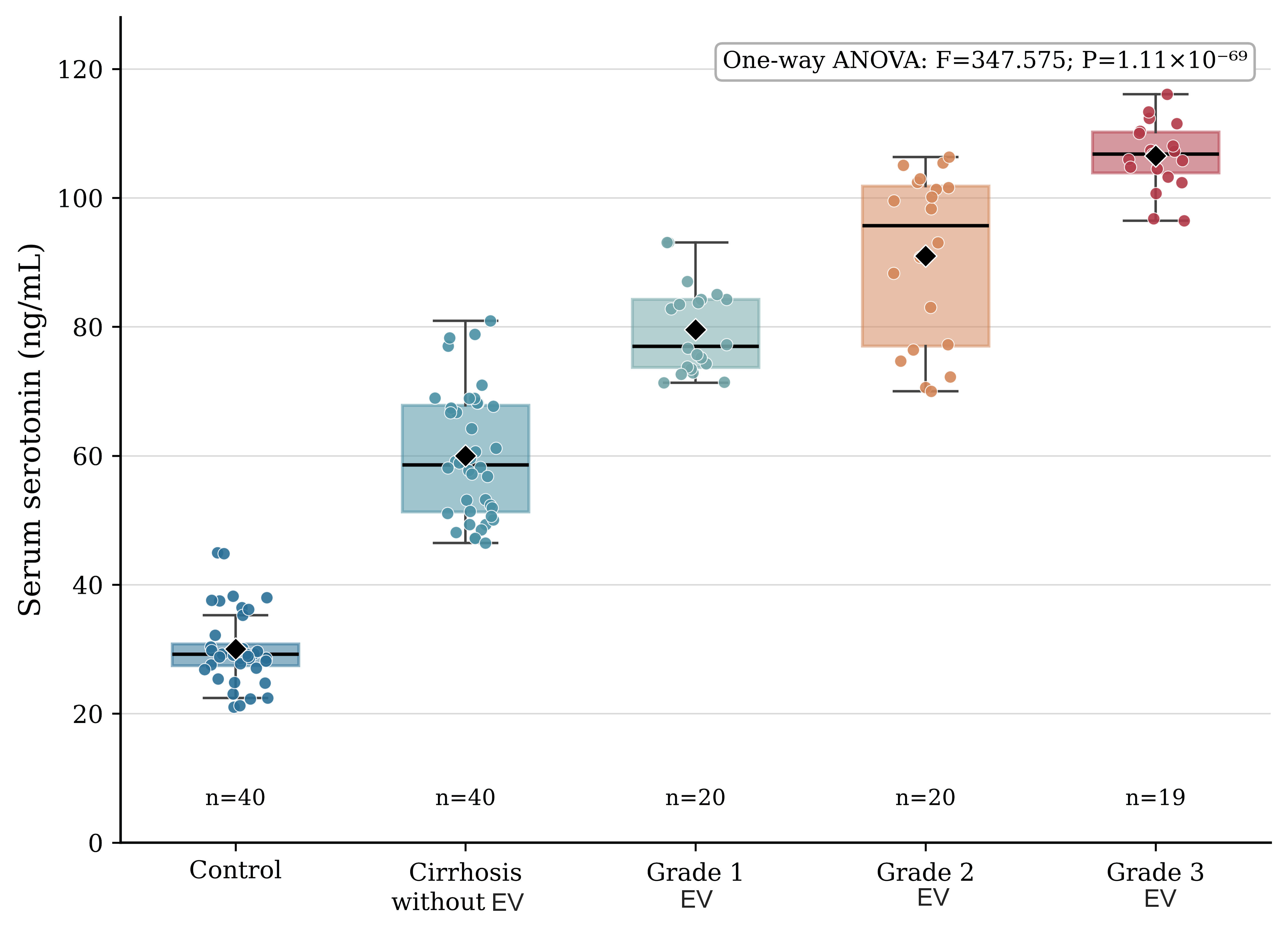 Click for large image | Figure 1. Serum serotonin concentrations across study groups. Box plots show median, interquartile range, and range; diamonds indicate group means; dots represent individual participants (n = 19 in the grade 3 group owing to one non-numeric (missing) serotonin value). ANOVA: analysis of variance; EV: esophageal varices. |
Ultrasonographic findings and composite portal hypertension indices
Liver size was normal in controls and shrunken with irregular nodular pattern across the four cirrhotic groups, consistent with progressive parenchymal shrinkage (H = 92.211, P < 0.001) (Table 3, Fig. 2). Portal vein diameter increased progressively with variceal grade, from 10.0 ± 0.7 mm in controls to 15.1 ± 0.4 mm in grade 3 varices (F = 190.412, P < 0.001), as did splenic vein diameter (F = 525.304, P < 0.001) and spleen diameter, from 8.6 cm in controls to a median of 15.3 cm in grade 3 varices (H = 102.260, P < 0.001). PSR fell correspondingly across the same groups, from 3,503.9 ± 482.8 in controls to 829.8 ± 553.0 in grade 3 varices (F = 63.593, P < 0.001). Increased liver echogenicity was near-universal across the cirrhotic groups but absent in controls (χ2 = 141.472, P < 0.001). Child-Pugh class C became progressively more frequent with increasing variceal grades, from 25.0% of participants with cirrhosis but no varices to 30.0% of grade 1, 60.0% of grade 2, and 70.0% of grade 3 participants.
Click to view | Table 3. Ultrasonographic Findings and Composite Portal Hypertension Indices |
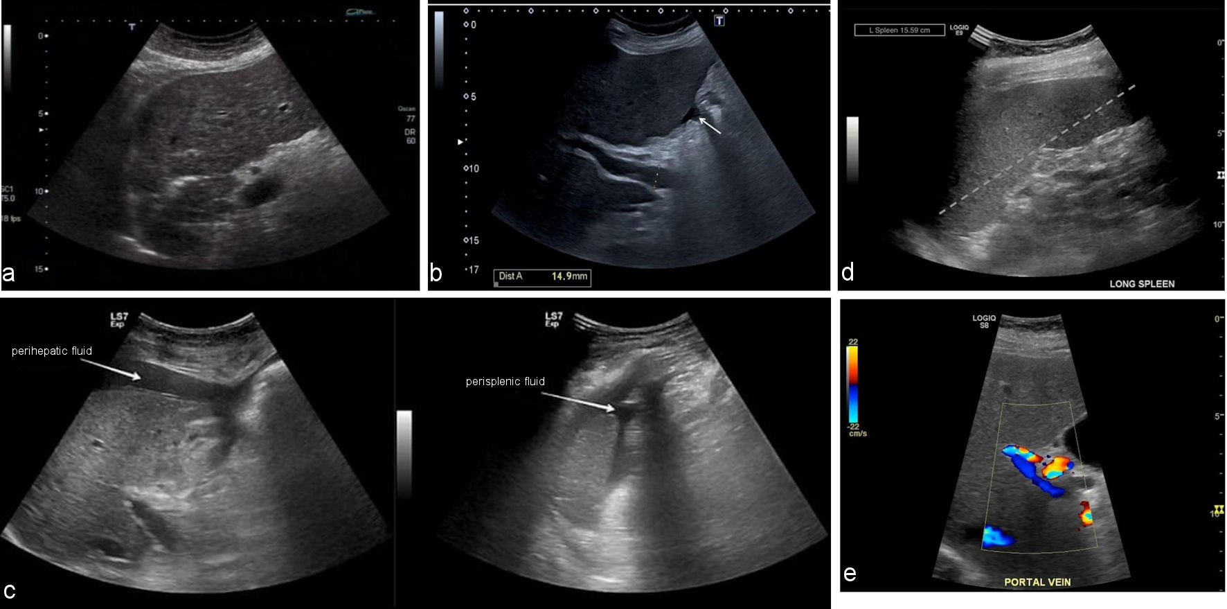 Click for large image | Figure 2. Representative abdominal ultrasound findings of cirrhosis and portal hypertension in the study cohort. (a) Greyscale ultrasound of the liver showing coarse, heterogeneous parenchymal echotexture with an irregular, nodular surface contour, typical of cirrhosis. (b) Greyscale ultrasound with caliper measurement of the main portal vein showing a diameter of 14.9 mm (arrow), exceeding the normal upper limit of 13 mm and consistent with portal hypertension. (c) Greyscale ultrasound demonstrating free perihepatic (left) and perisplenic (right) fluid collections (arrows), consistent with ascites. (d) Greyscale ultrasound of the long axis of the spleen showing splenomegaly, with a measured splenic length of 15.59 cm (dashed line), consistent with portal hypertension. (e) Color Doppler ultrasound of the main portal vein showing preserved hepatopetal flow within a dilated vessel. |
Predictors of esophageal variceal severity
Among the 60 participants with grade 1–3 varices, serum serotonin (ρ = 0.759, P < 0.001) and portal vein diameter (ρ = 0.754, P < 0.001) showed the strongest univariable correlation with variceal grade, followed by Child-Pugh class (ρ = 0.395, P = 0.002) and PSR (ρ = −0.381, P = 0.003) (Table 4). In the multivariable ordinal logistic regression model (n = 59 with complete serotonin data; likelihood-ratio χ2 = 77.882, df = 6, P < 0.001; McFadden pseudo-R2 = 0.601), serum serotonin (adjusted odds ratio (OR) of 7.48 per 10 ng/mL increase; 95% CI, 2.29–24.41; P = 0.0009) and portal vein diameter (adjusted OR of 1.70 per 0.1 mm increase; 95% CI, 1.29–2.23, P = 0.0002; equivalent to 197.1 per 1 mm, 95% CI, 12.7–3059.4) each remained independently and significantly associated with higher variceal grade after mutual adjustment. Child-Pugh class, PSR, age, and sex did not reach independent statistical significance once serotonin and portal vein diameter were accounted for. The very wide CI of the per-millimeter portal vein estimate reflects the narrow range of portal vein diameter among participants with varices (group means 14.0–15.1 mm) and the small sample relative to the number of model parameters; the unpenalized estimates should therefore be regarded as imprecise. In the Firth-penalized model, serotonin (adjusted OR of 5.68 per 10 ng/mL; 95% CI, 2.19–14.70; P = 0.0003) and portal vein diameter (adjusted OR of 1.56 per 0.1 mm; 95% CI, 1.24–1.96; P = 0.0001) remained the only independent predictors, as they did in the parsimonious model containing these two variables alone (OR = 4.19; 95% CI, 1.87–9.41; P = 0.0005; and OR = 1.53; 95% CI 1.24–1.89; P = 0.0001, respectively). Across 1,000 bootstrap resamples of the full model, the ORs for serotonin and portal vein diameter exceeded 1 in every resample, although about 5% of resamples produced extreme estimates consistent with quasi-separation; the direction of both associations was therefore robust, whereas their unpenalized magnitude was not.
Click to view | Table 4. Univariable Correlation and Multivariable Ordinal Regression Analysis of Predictors of Esophageal Variceal Severity (N = 59–60, Participants With Grade 1–3 Varices) |
Diagnostic performance for high-grade varices
Among the 99 cirrhotic participants with a complete serotonin measurement, 39 had grade 2 or 3 varices (Table 5, Fig. 3). The apparent AUC for high-grade varices was 0.944 (95% CI, 0.895–0.980) for serum serotonin, compared with portal vein diameter (AUC 0.923; 95% CI, 0.864–0.974), PSR (AUC 0.764; 95% CI, 0.660–0.861), and Child-Pugh class (AUC 0.746; 95% CI, 0.654–0.830). On DeLong’s test, the AUCs of serotonin and portal vein diameter did not differ significantly (difference 0.021; 95% CI, −0.049 to 0.091; P = 0.553), whereas serotonin exceeded both PSR (difference 0.180; 95% CI, 0.093–0.268; P < 0.001) and Child-Pugh class (difference 0.198; 95% CI, 0.112–0.285; P < 0.001); portal vein diameter likewise exceeded PSR (P = 0.004) and Child-Pugh class (P < 0.001), and PSR and Child-Pugh class did not differ (P = 0.687). At its Youden-optimal cutoff of 88.3 ng/mL or higher, serotonin showed an apparent sensitivity of 82.1% (95% CI, 67.3–91.0) and specificity of 96.7% (95% CI, 88.6–99.1), corresponding to a positive likelihood ratio of 24.6 and a negative likelihood ratio of 0.19 (Table 6). Its positive and negative predictive values of 94.1% and 89.2% reflect the 39.4% prevalence of high-grade varices fixed by the study design; at illustrative prevalences of 10% and 25%, the corresponding values would be 73.2% and 98.0%, and 89.1% and 94.2%, respectively (Table 6). After bootstrap optimism correction, the sensitivity and specificity of the serotonin cutoff were 80.1% and 95.9% (leave-one-out cross-validated, 79.5% and 96.7%), and corrections for the other markers were similarly small (Table 6). A post-hoc logistic model combining all four markers reached an apparent AUC of 0.984 in this same sample (0.979 after bootstrap optimism correction); because this combined model was neither derived nor tested in an independent cohort, its performance is reported as exploratory only and should not be interpreted as validated diagnostic accuracy. None of the four individual markers reached this level of apparent accuracy alone, which is consistent with each capturing a partially distinct aspect of portal hypertensive pathophysiology; the practical implication is that no single noninvasive test, on the evidence presented here, is likely to fully replace endoscopic assessment in an individual patient.
Click to view | Table 5. Diagnostic Performance (ROC Analysis) of Noninvasive Markers for Identifying Grade 2 or 3 Esophageal Varices |
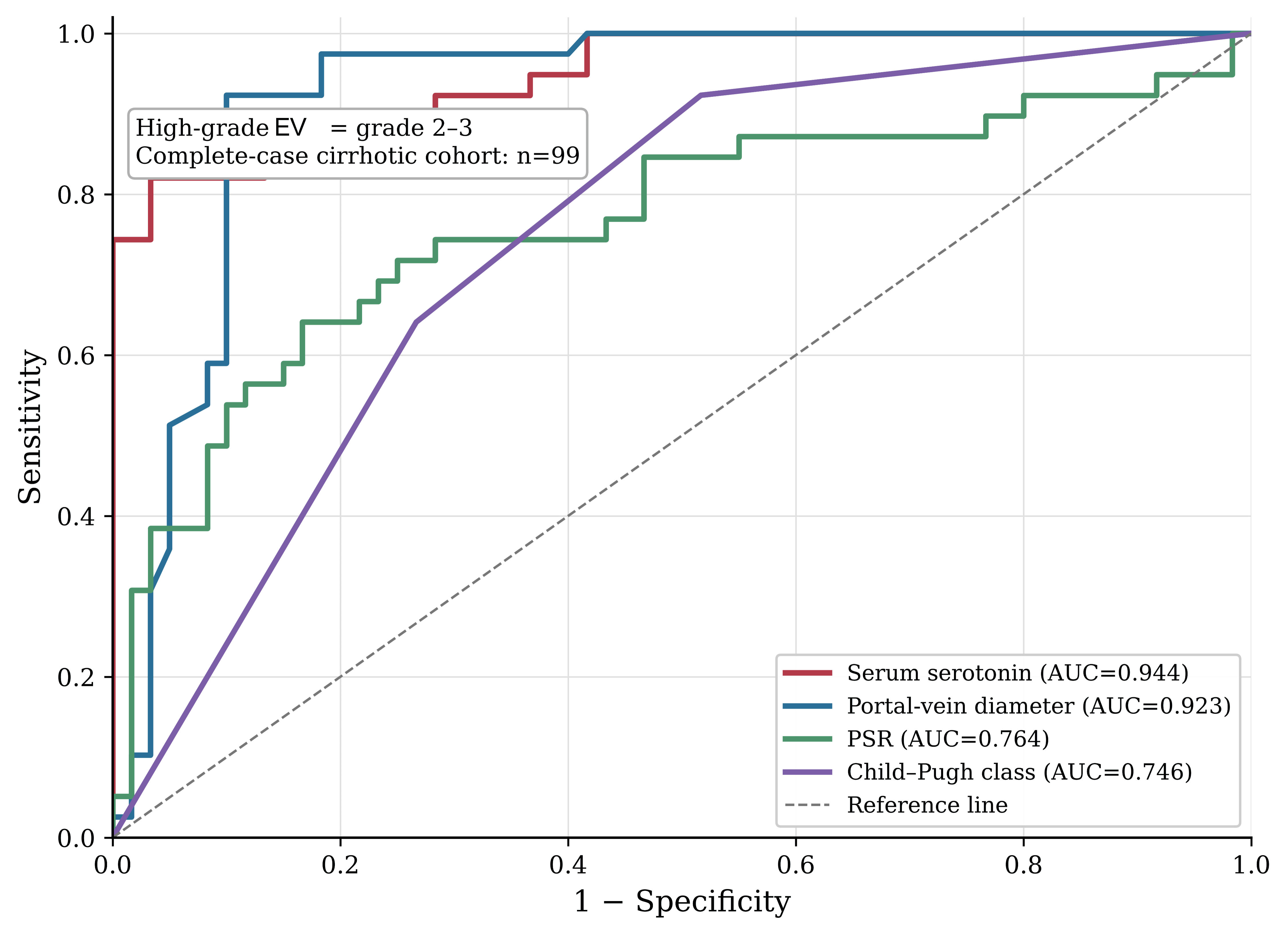 Click for large image | Figure 3. Receiver operating characteristic curves for noninvasive markers of high-grade esophageal varices. The diagonal dashed line represents the line of non-discrimination (AUC = 0.5). EV: esophageal varices; AUC: area under the curve; PSR: platelet count to spleen diameter ratio. |
Click to view | Table 6. Exploratory Accuracy Measures for Identifying Grade 2 or 3 Esophageal Varices: Optimism-Corrected, Prevalence-Independent, and Prevalence-Adjusted Estimates |
| Discussion | ▴Top |
In this cross-sectional study of 140 participants spanning the full spectrum from the absence of liver disease to grade 3 esophageal varices, serum serotonin rose progressively with variceal severity and, together with portal vein diameter, remained an independent predictor of variceal grade after adjustment for Child-Pugh class, PSR, age, and sex. Serotonin and portal vein diameter showed similarly high discrimination for high-grade varices, with no significant difference between them on paired comparison, and in this enriched sample both discriminated significantly better than PSR and Child-Pugh class. These findings are consistent with the hypothesis that platelet-derived serotonin reflects, at least in part, the severity of the portal hypertensive process underlying variceal formation, rather than serving only as an incidental marker of hepatic dysfunction. The graded increase in serum serotonin across variceal grades was not reported in an earlier analysis of this cohort [18]; the per-grade values presented here were derived from the verified participant-level dataset (Table 2).
Because more than 90% of circulating serotonin is stored within platelet dense granules, serum concentration is shaped jointly by platelet turnover, splenic sequestration, and hepatic vascular handling [10]. Platelet-derived serotonin supports liver regeneration in humans [11] and modulates hepatic stellate cell activation, a central step in fibrogenesis [12], and blockade of its receptors has attenuated fibrosis in experimental models [13]. In the context of progressive portal hypertension, increased splenic platelet turnover and altered hepatic serotonin clearance could plausibly raise circulating concentrations in parallel with worsening variceal disease; however, the cross-sectional design of the present study cannot establish this mechanism directly, and the association observed here should be interpreted as correlative rather than causal. A further consideration is that serotonin is metabolized hepatically and renally, so impaired clearance in more advanced liver disease could, in principle, contribute to the rise in circulating concentration independently of platelet biology; distinguishing production, storage-release, and clearance contributions to serum serotonin was outside the scope of this cross-sectional design.
PSR and portal vein diameter both moved in the expected direction with variceal severity, consistent with previous studies [7–9], but PSR did not remain an independent predictor once serotonin and portal vein diameter were included in the multivariable model, and its univariable correlation with grade was weaker than that of either serotonin or portal vein diameter. This pattern is compatible with PSR capturing much of the same underlying hypersplenism and portal-pressure information that is already reflected, separately, in spleen diameter and platelet count, rather than adding independent discriminative value once those pathways are represented by a direct anatomical measurement. Portal vein diameter, despite its relative simplicity, performed strongly in this cohort, though ultrasonographic measurement is more operator-dependent than a laboratory assay, a difference that should temper any direct comparison of the two markers’ apparent accuracy.
If replicated in independent, unselected surveillance cohorts, a noninvasive assessment anchored on serum serotonin and portal vein diameter could help identify patients most likely to have high-grade varices before endoscopy is performed, consistent with the broader shift toward risk-stratified, rather than universal, endoscopic screening reflected in current consensus and practice guidance [3, 17]. Because acute variceal hemorrhage is an important precipitant of hepatic encephalopathy and can necessitate urgent escalation of care in decompensated cirrhosis [14], earlier noninvasive identification of patients at highest risk could support more timely surveillance and prophylactic treatment, potentially reducing both the burden of unnecessary endoscopy among low-risk patients and the risk of delayed diagnosis among those at genuine risk of bleeding. Any such pathway would need to be validated against hard clinical endpoints, such as incident variceal hemorrhage on follow-up, rather than against endoscopic grade alone, before it could reasonably influence surveillance intervals in practice.
Strengths of this study include the simultaneous evaluation of a novel biochemical marker alongside three established noninvasive indices within the same cohort, endoscopic confirmation of the primary outcome in every participant, a five-group design spanning the full disease spectrum from non-hepatic controls to grade 3 varices, and a statistically prespecified, distribution-driven choice of test for each variable rather than a single default method applied uniformly across a heterogeneous set of clinical and laboratory measures.
This study also has several limitations. Its cross-sectional design cannot establish whether serotonin elevation precedes or follows variceal progression, and a longitudinal cohort with serial endoscopic and biochemical assessment would be needed to test this directly [25]. Participants were recruited into strata of prespecified size, so the distribution of variceal grades was fixed by design and enriched for high-grade varices relative to an unselected surveillance population; together with the comparison of clearly separated disease categories, this spectrum effect is likely to have inflated the apparent discriminative performance of all four markers [26]. The modest per-group sample sizes, particularly for the multivariable model (n = 59), limit precision and generalizability, and the wide CI around the portal vein diameter OR reflect this constraint. The multivariable model estimated six parameters from 59 participants, fewer than commonly recommended for multivariable modeling [20]; although the two principal associations persisted in penalized and parsimonious models and were directionally consistent across bootstrap resamples, their magnitude remains imprecise and should be confirmed in larger cohorts. Serum serotonin is sensitive to pre-analytical handling and platelet degranulation, and although a standardized collection protocol was followed, some residual measurement variability cannot be excluded; moreover, serum rather than platelet-poor plasma serotonin was measured and values were not normalized to platelet count, so the contributions of platelet serotonin content and free circulating serotonin could not be separated. Serum was stored at −20 °C rather than −80 °C, the interval between collection and centrifugation and the number of freeze-thaw cycles were not recorded, and assay precision was not re-verified in-house. Blinding of the laboratory staff to group allocation was not documented, and variceal grading relied on the real-time assessment of the performing endoscopist without a documented independent second reading. The optimal cutoffs were derived in the same sample; bootstrap optimism correction and cross-validation reduced the apparent estimates only modestly, but internal validation cannot substitute for external validation, and predictive values depend on the prevalence fixed by the design. Finally, because the exploratory model combining all four markers was developed and evaluated in the same sample, its apparent AUC of 0.984 should not be interpreted as validated external diagnostic performance, and this figure is not presented as a clinical decision threshold.
Conclusions
In this cohort spanning non-hepatic controls, cirrhosis without varices, and grade 1 to grade 3 esophageal varices, serum serotonin increased progressively with variceal severity and, together with portal vein diameter, was independently associated with higher variceal grade after adjustment for Child-Pugh class, PSR, age, and sex. Serotonin discriminated grade 2 or 3 varices as well as portal vein diameter, with no significant difference between the two, and better than PSR and Child-Pugh class; however, these diagnostic estimates are exploratory, were obtained in a sample enriched by design, and require external validation. These findings support serum serotonin as a biologically plausible, measurable correlate of variceal severity that may complement, rather than replace, established noninvasive indices when risk-stratifying patients with cirrhosis. Prospective, multicenter studies using pre-analytically standardized serotonin assays and, ideally, longitudinal endoscopic follow-up are needed before serum serotonin, alone or in combination with portal vein diameter, can be recommended as a tool to guide the timing and frequency of endoscopic variceal surveillance.
Acknowledgments
None to declare.
Financial Disclosure
No external funding was received for this study.
Conflict of Interest
The authors declared no conflict of interest relevant to the content of the manuscript.
Informed Consent
Written informed consent was obtained from each participant before enrollment.
Author Contributions
AMF and AME contributed equally to this work and shared first authorship. AMF and AME conceived and designed the study, performed the upper gastrointestinal endoscopies and variceal grading, and wrote the first draft of the manuscript. AAk supervised the laboratory work, including the serum serotonin ELISA and routine laboratory investigations, and contributed to the data analysis and interpretation. ASEE performed and interpreted the abdominal ultrasound and Doppler examinations. AEE, FSM, NRM, OMAE, AAMA, AAb, MAM, IAMIB, and AAr recruited patients, carried out the clinical assessment and Child-Pugh scoring, and collected the data. SASA performed the statistical analysis and contributed to the interpretation of the data. SMEN supervised the study and critically revised the manuscript for important intellectual content. All authors read and approved the final manuscript and agree to be accountable for all aspects of the work.
Data Availability
The datasets generated and analyzed during the current study are available from the corresponding author on reasonable request.
AI Use Declaration
During the preparation of this manuscript, the authors used AI to assist with language editing and with formatting the manuscript, tables, and references according to the journal’s author guidelines. The tool was not used to generate data, perform or interpret statistical analyses, or draw scientific conclusions. The authors reviewed and edited all output and took full responsibility for the content, citations, analyses, interpretations, and conclusions of this publication. No AI tool is listed as an author.
Abbreviations
ALP: alkaline phosphatase; ALT: alanine aminotransferase; ANOVA: analysis of variance; AST: aspartate aminotransferase; AUC: area under the receiver operating characteristic curve; CI: confidence interval; ELISA: enzyme-linked immunosorbent assay; FFH: Fisher–Freeman–Halton; INR: international normalized ratio; IQR: interquartile range; IRB: institutional review board; LR+/LR−: positive/negative likelihood ratio; NPV: negative predictive value; OR: odds ratio; EV: esophageal varices; PPV: positive predictive value; PSR: platelet count to spleen diameter ratio; ROC: receiver operating characteristic; SD: standard deviation; STROBE: Strengthening the Reporting of Observational Studies in Epidemiology
| References | ▴Top |
- G. B. D. Cirrhosis Collaborators. The global, regional, and national burden of cirrhosis by cause in 195 countries and territories, 1990-2017: a systematic analysis for the Global Burden of Disease Study 2017. Lancet Gastroenterol Hepatol. 2020;5(3):245-266.
doi pubmed - Moon AM, Singal AG, Tapper EB. Contemporary epidemiology of chronic liver disease and cirrhosis. Clin Gastroenterol Hepatol. 2020;18(12):2650-2666.
doi pubmed - de Franchis R, Bosch J, Garcia-Tsao G, Reiberger T, Ripoll C, Baveno VIIF. Baveno VII - Renewing consensus in portal hypertension. J Hepatol. 2022;76(4):959-974.
doi pubmed - Ruf A, Dirchwolf M, Freeman RB. From Child-Pugh to MELD score and beyond: Taking a walk down memory lane. Ann Hepatol. 2022;27(1):100535.
doi pubmed - Kotak PS, Kumar J, Kumar S, Varma A, Acharya S. Navigating cirrhosis: a comprehensive review of liver scoring systems for diagnosis and prognosis. Cureus. 2024;16(3):e57162.
doi pubmed - Katwal S, Ansari MA, Suwal S, Rayamajhi S, Ghimire P, Ghimire A. Measurement of portal vein indices and splenic index by ultrasound and their association with gastroesophageal varices in cirrhosis of liver. Ann Med Surg (Lond). 2023;85(12):5926-5931.
doi pubmed - Mossie GY, Nur AM, Ayalew ZS, Azibte GT, Berhane KA. Platelet counts to spleen diameter ratio: A promising noninvasive tool for predicting esophageal varices in cirrhosis patients. World J Hepatol. 2024;16(10):1177-1187.
doi pubmed - Rapelly SS, Singh S, Verma N, Bhattacharya S, Rungta S. Non-invasive predictors to grade esophageal varices in liver cirrhosis patients. J Family Med Prim Care. 2024;13(4):1232-1237.
doi pubmed - Eldesoky AE, Aeriba M, Abd ElFattah F, Elhawary G, Awad M. Platelet count/spleen diameter ratio as a predictor of high-risk esophageal varices in patients with liver cirrhosis. Med J Viral Hepat. 2022;6(2):25-30.
- Belmer A, Luci C, Gual P. Role of central and peripheral serotonin in liver physiology and diseases. Theranostics. 2026;16(5):2284-2309.
doi pubmed - Starlinger P, Pereyra D, Hackl H, Ortmayr G, Braunwarth E, Santol J, Najarnia S, et al. Consequences of perioperative serotonin reuptake inhibitor treatment during hepatic surgery. Hepatology. 2021;73(5):1956-1966.
doi pubmed - Redensek Trampuz S, van Riet S, Nordling A, Ingelman-Sundberg M. Mechanisms of 5-HT receptor antagonists in the regulation of fibrosis in a 3D human liver spheroid model. Sci Rep. 2024;14(1):1396.
doi pubmed - Zhou XY, Xu Z, Liu X, Wu G, Hou Y, Qi LW, Liu Q, et al. Bifunctional LYTAC mediates hepatocytes-hepatic stellate cells crosstalk by regulating 5-HT(2A) receptor degradation and antagonism to synergistically ameliorate hepatic fibrosis. J Am Chem Soc. 2025;147(48):44162-44174.
doi pubmed - European Association for the Study of the Liver. EASL Clinical Practice Guidelines on the management of hepatic encephalopathy. J Hepatol. 2022;77(3):807-824.
doi pubmed - World Medical Association. World Medical Association Declaration of Helsinki: Ethical Principles for Medical Research Involving Human Participants. JAMA. 2025;333(1):71-74.
doi pubmed - von Elm E, Altman DG, Egger M, Pocock SJ, Gotzsche PC, Vandenbroucke JP, Initiative S. The Strengthening the Reporting of Observational Studies in Epidemiology (STROBE) statement: guidelines for reporting observational studies. Lancet. 2007;370(9596):1453-1457.
doi pubmed - Kaplan DE, Ripoll C, Thiele M, Fortune BE, Simonetto DA, Garcia-Tsao G, Bosch J. AASLD Practice Guidance on risk stratification and management of portal hypertension and varices in cirrhosis. Hepatology. 2024;79(5):1180-1211.
doi pubmed - Abd El-Atey MA, Bastawy MB, Abd El-Razek OM, Ali AA. The correlation between serum serotonin level and grade of esophageal varices in patients with liver cirrhosis. Int J Med Arts. 2025.
doi - Liu A, He H, Tu XM, Tang W. On testing proportional odds assumptions for proportional odds models. Gen Psychiatr. 2023;36(3):e101048.
doi pubmed - Riley RD, Ensor J, Snell KIE, Harrell FE, Jr., Martin GP, Reitsma JB, Moons KGM, et al. Calculating the sample size required for developing a clinical prediction model. BMJ. 2020;368:m441.
doi pubmed - Suhas S, Manjunatha N, Kumar CN, Benegal V, Rao GN, Varghese M, Gururaj G. Firth's penalized logistic regression: a superior approach for analysis of data from India's National Mental Health Survey, 2016. Indian J Psychiatry. 2023;65(12):1208-1213.
doi pubmed - Hassanzad M, Hajian-Tilaki K. Methods of determining optimal cut-point of diagnostic biomarkers with application of clinical data in ROC analysis: an update review. BMC Med Res Methodol. 2024;24(1):84.
doi pubmed - Efthimiou O, Seo M, Chalkou K, Debray T, Egger M, Salanti G. Developing clinical prediction models: a step-by-step guide. BMJ. 2024;386:e078276.
doi pubmed - Nahm FS. Receiver operating characteristic curve: overview and practical use for clinicians. Korean J Anesthesiol. 2022;75(1):25-36.
doi pubmed - Gines P, Krag A, Abraldes JG, Sola E, Fabrellas N, Kamath PS. Liver cirrhosis. Lancet. 2021;398(10308):1359-1376.
doi pubmed - Pavlou A, Kurtz RM, Song JW. Diagnostic accuracy studies in radiology: How to recognize and address potential sources of bias. Radiol Res Pract. 2021;2021:5801662.
doi pubmed
This article is distributed under the terms of the Creative Commons Attribution 4.0 International License (CC BY 4.0), which permits unrestricted use, distribution, and reproduction in any medium, including commercial use, provided the original work is properly cited.
Journal of Clinical Medicine Research is published by Elmer Press Inc.