Spatial SRY-Box Transcription Factor 2 Expression and Stemness-Associated Markers Refine Risk Stratification in Barrett’s Esophagus: A Digital Pathology Study of 217 Patients
DOI:
https://doi.org/10.14740/jocmr6562Keywords:
SOX2, Barrett’s esophagus, esophageal adenocarcinoma, spatial heterogeneity, digital pathology, risk stratificationAbstract
Background: Barrett’s esophagus (BE) is a precursor lesion of esophageal adenocarcinoma (EAC), but current risk stratification remains largely dependent on dysplasia grade and segment length. We investigated whether spatial SRY-box transcription factor 2 (SOX2) expression patterns could improve prediction of neoplastic progression in BE.
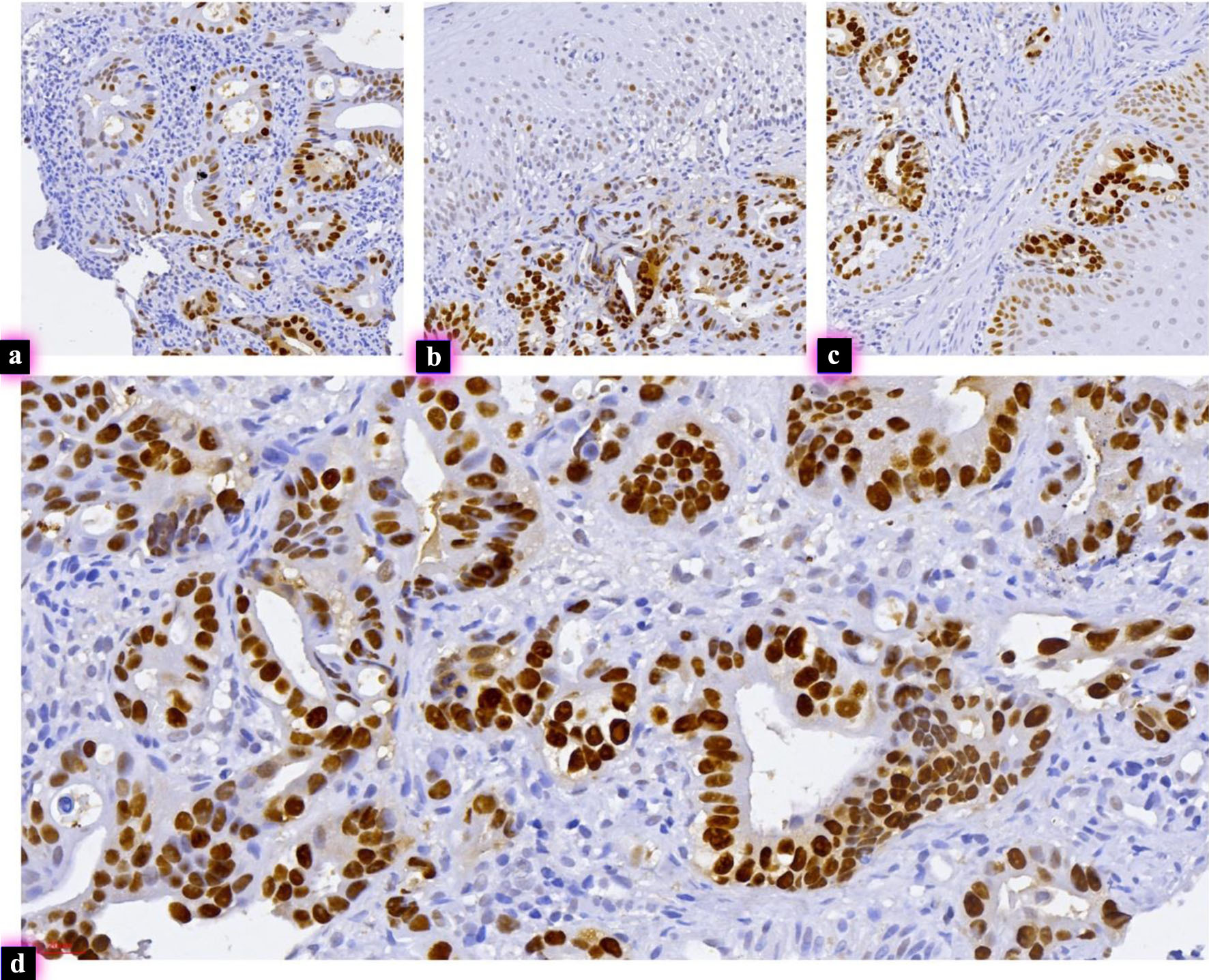
Methods: This retrospective multicenter study included 217 patients with reflux esophagitis, non-dysplastic BE (NDBE), low-grade dysplasia (LGD), high-grade dysplasia (HGD), or intramucosal EAC (EAC_T1a). Immunohistochemistry for SOX2, cluster of differentiation 44 (CD44), E-cadherin, p53, Ki-67 proliferation antigen (Ki67), and caudal-type homeobox transcription factor 2 (CDX2) was performed on formalin-fixed paraffin-embedded tissue. SOX2 expression was evaluated at the squamo–columnar junction and in deep Barrett glands, and a SOX2 gradient was calculated. Digital image analysis was used to assess SOX2 spatial heterogeneity and SOX2/CD44 co-localization. Associations with progression were analyzed using non-parametric tests and logistic regression models.
Results: Progressive dysplastic transformation was associated with inversion of the SOX2 gradient, increased SOX2 spatial heterogeneity, and expansion of SOX2/CD44 double-positive glands. Median SOX2_Gradient shifted from positive values in reflux esophagitis and NDBE to strongly negative values in HGD and EAC_T1a (P < 0.001). SOX2_Tile_Gini and CD44_HScore increased significantly with histologic severity and progression status (both, P < 0.001). Lower SOX2_Gradient, higher SOX2_Tile_Gini, higher CD44_HScore, and longer Prague M length were significantly associated with progression to HGD/EAC.
Conclusions: Spatial remodeling of SOX2 expression is strongly associated with dysplasia grade and neoplastic progression in BE. Integration of SOX2 spatial metrics with established biomarkers may improve risk stratification beyond conventional histopathologic assessment.
Published
Issue
Section
License
Copyright (c) 2026 The authors

This work is licensed under a Creative Commons Attribution 4.0 International License.