Quantitative Facet Joint Effusion on Magnetic Resonance Imaging Is Associated With Dynamic Segmental Instability and Pain Severity in Degenerative Lumbar Spondylolisthesis
DOI:
https://doi.org/10.14740/jocmr6572Keywords:
Facet joint effusion, Facet joint degeneration, MRI, Lumbar spine instability, Degenerative lumbar spondylolisthesisAbstract
Background: The aims of the study were to determine whether quantitatively measured facet joint effusion (FJE) on magnetic resonance imaging (MRI) is associated with dynamic segmental instability and greater pain severity in patients with degenerative lumbar spondylolisthesis (DLS), and to evaluate the diagnostic performance of FJE for identifying unstable segments.
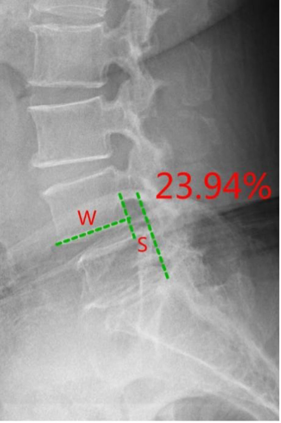
Methods: This retrospective single-center study reviewed 151 consecutive DLS patients treated at Qilu Hospital (Qingdao) between January 1, 2020 and June 30, 2024. The cohort included 31 men and 118 women (mean age 64.52 ± 9.76 years). Slipped levels comprised L3 (n = 7), L4 (n = 116), and L5 (n = 28). Patients with prior lumbar surgery, acute spinal trauma, tumor, ankylosing spondylitis, marked scoliosis, multilevel spondylolisthesis, or retrolisthesis were excluded. On axial T2-weighted MRI, the maximal unilateral and bilateral facet effusion thicknesses were measured in millimeters. Dynamic imaging was used to classify segments as stable or unstable. Pain was quantified using the Visual Analog Scale (VAS). Statistical analyses compared FJE presence and size between stable and unstable segments and assessed diagnostic accuracy (sensitivity, specificity, receiver operating characteristic area under the curve) and correlations with slip distance and VAS.
Results: Seventy-two patients (47.7%) had dynamic segmental instability. FJE was present at 93.1% of unstable levels, with mean effusion thickness of 2.75 ± 0.89 mm, whereas stable levels showed a 26.6% FJE incidence and mean thickness of 1.25 ± 1.25 mm (P < 0.05). Using presence of FJE to identify instability yielded sensitivity 93.1% and specificity 73.4%. Among 88 patients with effusion ≥ 1.0 mm, 76.1% exhibited instability. Effusion thickness correlated linearly with the anteroposterior slip distance difference. Facet joint degeneration grade related nonlinearly to effusion width (increase from grade 1 to 2, decline at grade 3; P < 0.05). ROC AUCs for left and right effusion thickness were 0.9243 and 0.9296, respectively (P < 0.0001). Patients with effusion had higher VAS scores (4.30 ± 1.07 vs. 2.79 ± 0.68, P < 0.05).
Conclusions: Millimeter-quantified FJE on MRI was strongly associated with dynamic segmental instability and greater pain severity in DLS. Quantitative FJE demonstrated high diagnostic accuracy and may serve as a practical imaging marker to identify unstable lumbar segments. Given the retrospective design, these results indicate association rather than causation and require prospective validation.
Published
Issue
Section
License
Copyright (c) 2026 The authors

This work is licensed under a Creative Commons Attribution 4.0 International License.