Fetus-in-Fetu: A PRISMA-Informed Narrative Review With an Illustrative Prenatal Case
DOI:
https://doi.org/10.14740/jocmr6423Keywords:
Fetus-in-fetu, Teratoma, Embryology, Congenital abnormalities, Prenatal diagnosis, Neonatal surgery, Parasitic twinAbstract
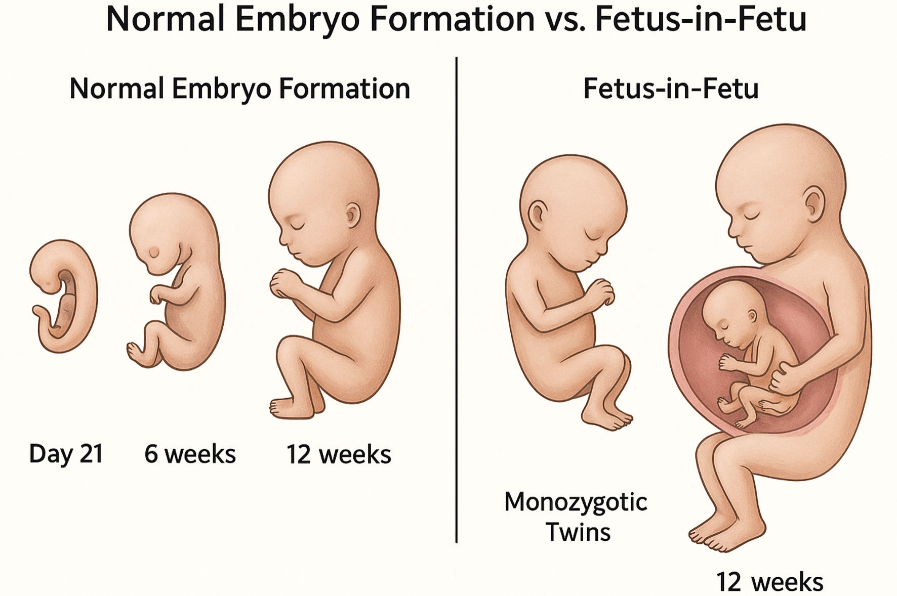
Fetus-in-fetu (FIF) is an uncommon congenital anomaly arising from aberrant monozygotic twinning, in which a malformed parasitic twin becomes incorporated within the body of the host. Despite its rarity, FIF remains clinically relevant because it is frequently misdiagnosed as a teratoma, particularly when detected outside the neonatal period or at atypical anatomical sites. This review present a Preferred Reporting Items for Systematic Reviews and Meta-Analyses (PRISMA)-informed narrative synthesis of the human literature on FIF, integrating embryological concepts, diagnostic imaging features, genetic observations, and surgical management. A systematic search of major electronic databases identified 45 publications meeting predefined eligibility criteria. From these, 25 core studies were selected for in-depth narrative analysis based on diagnostic confirmation, methodological clarity, and avoidance of overlapping case reports. To contextualize the literature, a previously unreported human case of FIF associated with omphalocele and complex congenital cardiac anomalies was also described, illustrating current prenatal diagnostic pathways and multidisciplinary postnatal care. Across reported cases, FIF is most often diagnosed in infancy and is typically benign following complete surgical excision. Distinguishing FIF from mature teratoma relies on recognition of organized axial structures, symmetry, and shared monozygotic genetic identity. Although malignant transformation is rare, incomplete resection and immature tissue components warrant long-term surveillance. Improved prenatal imaging has enhanced early recognition of FIF, yet its developmental mechanisms remain incompletely understood. Future progress will likely depend on coordinated clinical reporting and translational research addressing early embryonic asymmetry, diagnostic refinement, and evidence-based follow-up strategies.
Published
Issue
Section
License
Copyright (c) 2026 The authors

This work is licensed under a Creative Commons Attribution 4.0 International License.