

When Stress Biology Meets Coronary Atherosclerosis: Diurnal Cortisol Patterns and Coronary Artery Disease Burden in Patients With Diabetes and Chronic Depression
DOI:
https://doi.org/10.14740/jocmr6749Keywords:
Coronary artery disease, Diurnal cortisol, Type 2 diabetes mellitus, Chronic depression, Hypothalamic–pituitary–adrenal axis, Coronary angiography, Stress biomarkersAbstract
Background: Type 2 diabetes mellitus and chronic depression frequently co-occur and jointly accelerate coronary artery disease (CAD), and dysregulation of the hypothalamic–pituitary–adrenal axis has been proposed as a biological link. This study aimed to determine whether diurnal serum cortisol metrics are associated with the angiographic burden of CAD in patients with both conditions.
Methods: In this retrospective observational study with a case–control component, 100 participants were analyzed: 50 patients with type 2 diabetes and clinician-documented chronic depression (cases) and 50 healthy controls. Morning (06:00–10:00) and evening (16:00–20:00) serum cortisol was measured with the Roche Elecsys Cortisol II immunoassay, and the AM–PM difference and AM/PM ratio were derived. In cases, coronary burden was classified from catheter angiography reports as multivessel CAD (≥ 70% stenosis in two or more major epicardial arteries) and obstructive CAD (≥ 70% stenosis in any epicardial vessel or ≥ 50% in the left main). Group comparisons, exploratory multivariable logistic regression adjusted for age, sex, body mass index, hypertension, dyslipidemia and smoking, and receiver operating characteristic (ROC) analyses were performed; because of the small number of events, the adjusted and ROC analyses are reported as exploratory and hypothesis-generating.
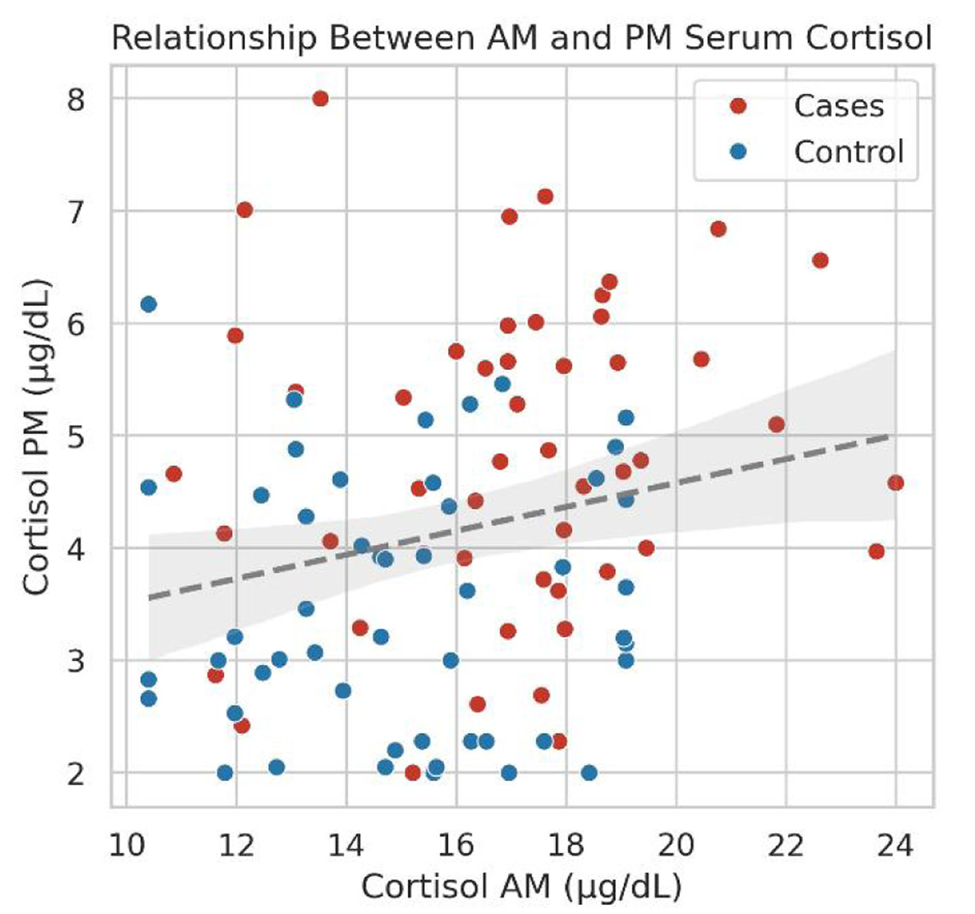
Results: Compared with controls, cases had higher morning cortisol (17.00 ± 3.00 vs. 15.02 ± 2.63 µg/dL; mean difference 1.98 µg/dL, 95% confidence interval (CI): 0.86–3.10, P = 0.0007), higher evening cortisol (median 4.72 vs. 3.21 µg/dL, P < 0.001), and a lower AM/PM ratio (median 3.56 vs. 4.28, P = 0.0087), whereas the absolute AM–PM difference did not differ (P = 0.25). Among cases, 37 had multivessel CAD and 19 had obstructive CAD. No cortisol parameter differed according to multivessel (all P ≥ 0.29) or obstructive status (all P ≥ 0.52), and, in exploratory adjusted models, none was independently associated with either endpoint (e.g., adjusted odds ratio (aOR) for evening cortisol 0.61, 95% CI 0.33–1.11 for multivessel CAD and 0.74, 95% CI 0.41–1.33 for obstructive CAD). In exploratory ROC analyses, cortisol metrics showed poor discrimination, with areas under the curve ranging from 0.39 (95% CI 0.22–0.59) to 0.59 (95% CI 0.43–0.74); every 95% CI included 0.50. In the exploratory adjusted model, older age was inversely associated with obstructive CAD (aOR 0.91, 95% CI 0.84–0.99, P = 0.030), a finding that requires confirmation given only 19 obstructive CAD events.
Conclusions: Patients with comorbid type 2 diabetes and chronic depression show elevated cortisol with a blunted diurnal ratio, but single-day spot serum cortisol metrics are not associated with the angiographic extent or severity of CAD in this sample; the adjusted and ROC analyses were exploratory and require confirmation. Measures of cumulative cortisol exposure, such as repeated multi-day salivary sampling and hair cortisol, combined with comprehensive coronary computed tomography angiography, merit evaluation in larger prospective cohorts.
Published
Issue
Section
License
Copyright (c) 2026 The authors

This work is licensed under a Creative Commons Attribution 4.0 International License.